A persistent dull ache in the right testicle represents one of the most concerning yet frequently misunderstood urological complaints. This discomfort can significantly impact quality of life, affecting everything from sleep patterns to physical activity levels. The complexity of testicular innervation and the intricate network of structures within the scrotal anatomy mean that even subtle pathological changes can manifest as noticeable discomfort.
Understanding the underlying mechanisms behind right-sided testicular pain requires appreciation of both local and systemic factors. The condition affects men across all age groups, with varying presentations depending on the underlying aetiology. Chronic orchialgia , the medical term for persistent testicular pain lasting more than three months, affects approximately 100,000 men annually in the United Kingdom alone. Early recognition and appropriate management of these conditions can prevent progression to more serious complications and preserve both reproductive function and overall wellbeing.
Epididymitis and epididymo-orchitis: primary inflammatory causes
Inflammatory conditions of the epididymis and testis represent the most common identifiable causes of unilateral testicular pain. These conditions typically develop through ascending infection from the urethra or haematogenous spread from distant infectious foci. The inflammation process creates localised tissue swelling and increased pressure within the confined space of the tunica albuginea, resulting in the characteristic dull, aching sensation that patients describe.
The clinical presentation often includes gradual onset of pain over several days, accompanied by scrotal swelling, erythema, and occasionally fever. The pain typically worsens with physical activity and may be relieved by scrotal elevation, a phenomenon known as Prehn’s sign . This distinctive feature helps differentiate inflammatory conditions from testicular torsion, where elevation typically provides no relief or may worsen the discomfort.
Chlamydia trachomatis and neisseria gonorrhoeae infections
Sexually transmitted infections account for the majority of epididymitis cases in men under 35 years of age. Chlamydia trachomatis represents the most frequently isolated organism, responsible for approximately 60% of cases in this demographic. The organism’s predilection for columnar epithelium makes the epididymal tubules particularly susceptible to infection and subsequent inflammatory response.
Neisseria gonorrhoeae infections often present with more acute symptoms, including purulent urethral discharge and dysuria preceding the testicular pain. The inflammatory response tends to be more intense, with marked scrotal swelling and erythema developing rapidly. Co-infection with both organisms occurs in approximately 20% of cases, necessitating dual antimicrobial therapy for optimal treatment outcomes.
Escherichia coli and enterococcus faecalis in older men
Men over 35 years typically develop epididymitis secondary to urinary tract pathogens, with Escherichia coli and Enterococcus faecalis being the most common causative organisms. These infections often occur in the context of underlying urological abnormalities, such as benign prostatic hyperplasia, urethral strictures, or bladder dysfunction.
The pathogenesis involves retrograde ascent of bacteria through the vas deferens, facilitated by increased intravesical pressure during voiding or instrumentation. Risk factors include recent urological procedures, indwelling catheters, and immunocompromised states. The clinical presentation may be more subtle than in younger men, with gradual onset of symptoms and less pronounced systemic signs of infection.
Mycoplasma genitalium and ureaplasma urealyticum complications
These atypical organisms have gained recognition as important causes of non-gonococcal urethritis and subsequent epididymitis. Mycoplasma genitalium demonstrates particular tropism for urogenital tissues and has been associated with treatment-resistant cases due to emerging antimicrobial resistance patterns.
Ureaplasma urealyticum infections often present with more indolent symptoms, making diagnosis challenging. The organism’s ability to persist in prostatic tissue can lead to recurrent episodes of epididymitis, particularly in men with concurrent prostatitis. Treatment requires specific antimicrobial agents, as standard β-lactam antibiotics are ineffective against these wall-deficient organisms.
Non-infectious epididymitis from amiodarone and behçet’s disease
Drug-induced epididymitis represents an underappreciated cause of testicular pain, with amiodarone being the most frequently implicated agent. The mechanism involves direct tissue toxicity and inflammatory response, typically occurring after several months of therapy. The condition is dose-dependent and usually resolves following drug discontinuation, though symptoms may persist for several weeks.
Behçet’s disease, a multisystem vasculitic condition, can affect the genitourinary system in approximately 15% of male patients. Epididymo-orchitis may be the presenting manifestation, characterised by recurrent episodes of unilateral or bilateral testicular pain and swelling. The diagnosis requires correlation with other systemic features, including oral ulceration, skin lesions, and ocular involvement.
Testicular torsion variants and ischaemic conditions
Testicular torsion and its variants represent urological emergencies requiring immediate recognition and intervention. While acute torsion presents with sudden, severe pain, chronic or intermittent torsion can manifest as persistent dull aching, particularly on the right side. The anatomical predisposition to torsion varies between individuals, with certain congenital anomalies significantly increasing the risk of both acute and chronic ischaemic events.
The pathophysiology involves compromise of testicular blood supply through twisting of the spermatic cord. Even partial obstruction of venous drainage can result in chronic congestion and the characteristic dull, aching pain. The condition requires high clinical suspicion, as delayed diagnosis can result in testicular atrophy and loss of function. Bilateral orchiopexy is typically recommended once the diagnosis is confirmed, as the anatomical predisposition usually affects both sides.
Intermittent testicular torsion and bell-clapper deformity
Intermittent testicular torsion occurs when incomplete rotation of the spermatic cord creates temporary ischaemia followed by spontaneous detorsion. This condition affects approximately 16% of men presenting with acute scrotal pain and can progress to complete torsion in up to 50% of cases if left untreated.
The bell-clapper deformity represents the most significant anatomical risk factor, occurring when the testis lacks proper attachment to the tunica vaginalis. This abnormality allows excessive mobility within the scrotal compartment, predisposing to torsion events. Men with this anatomy may experience recurrent episodes of testicular pain, particularly during periods of increased physical activity or following minor trauma.
Torsion of testicular appendages: hydatid of morgagni
The testicular appendices, embryological remnants of Müllerian and Wolffian ducts, can undergo torsion and necrosis, resulting in localised pain and inflammation. The hydatid of Morgagni , or appendix testis, is the most commonly affected structure, present in approximately 90% of males and located at the superior pole of the testis.
This condition typically affects prepubertal boys but can occur in adults, presenting with sudden onset of localised testicular pain. The classic “blue dot sign,” representing the necrotic appendage visible through the scrotal skin, is pathognomonic when present. Unlike testicular torsion, the pain is usually localised to the upper pole of the testis and may be associated with a small, tender nodule palpable in this region.
Chronic testicular ischaemia following previous torsion
Men with a history of testicular torsion may develop chronic pain syndromes related to ongoing ischaemic changes or testicular atrophy. The compromised blood supply can result in chronic inflammation and fibrosis, leading to persistent discomfort months or years after the initial event.
Chronic ischaemic orchialgia affects approximately 12% of men following testicular torsion, with symptoms including dull aching pain, testicular atrophy, and reduced fertility potential.
The condition may also develop following partial torsion events that went unrecognised or inadequately treated. Salvage orchiopexy performed more than 6 hours after symptom onset carries increased risk of chronic complications due to ischaemia-reperfusion injury and subsequent inflammatory cascades.
Varicocele-induced venous congestion and temperature elevation
Varicoceles represent abnormal dilatation of the pampiniform venous plexus within the spermatic cord, affecting approximately 15% of the general male population and 35% of men with primary infertility. The condition predominantly affects the left side due to anatomical differences in venous drainage, but right-sided or bilateral varicoceles can occur.
The pathophysiology involves venous congestion and retrograde blood flow, leading to increased intrascrotal temperature and chronic testicular hypoxia. These changes result in oxidative stress, hormonal imbalances, and inflammatory responses that manifest as chronic testicular discomfort. The pain typically worsens with prolonged standing or physical exertion and improves with recumbency.
Urological trauma and post-procedural complications
Testicular trauma can result from various mechanisms, including blunt force impact during sporting activities, motor vehicle accidents, or direct assault. The immediate injury may resolve, but chronic pain syndromes can develop due to several factors including haematoma formation, chronic inflammation, and development of adhesions between tissue planes. The right testicle may be particularly susceptible to certain types of trauma due to anatomical positioning and the dominant hand preference in most interpersonal violence situations.
Post-procedural complications following urological interventions can also manifest as chronic testicular pain. Procedures such as inguinal hernia repair, varicocelectomy, hydrocelectomy, or vasectomy can result in chronic pain syndromes through various mechanisms. Nerve entrapment, chronic inflammation, sperm granuloma formation, and adhesion development all contribute to ongoing discomfort. The incidence of chronic pain following these procedures ranges from 0.5% to 37%, depending on the specific intervention and surgical technique employed.
Iatrogenic injury to the genitofemoral nerve, lateral femoral cutaneous nerve, or ilioinguinal nerve during surgical procedures can result in chronic neuropathic pain patterns. These nerves provide sensory innervation to the scrotal and testicular region, and their dysfunction can create persistent dysaesthetic symptoms. The pain is typically described as burning, shooting, or tingling in nature, though it can occasionally present as a dull ache, particularly when combined with inflammatory responses at the surgical site.
Conservative management approaches include anti-inflammatory medications, neuropathic pain agents such as gabapentin or pregabalin, and physical therapy modalities. More invasive interventions may be considered for refractory cases, including nerve blocks, neurolysis procedures, or revision surgery to address anatomical causes of ongoing symptoms. The success rates of these interventions vary considerably, emphasising the importance of prevention through careful surgical technique and appropriate patient counselling regarding potential complications.
Neoplastic conditions: seminomas and non-seminomatous germ cell tumours
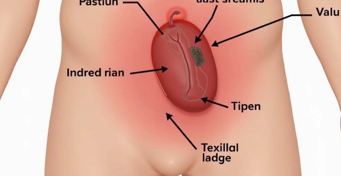
Testicular malignancies, while relatively uncommon, represent the most serious potential cause of testicular pain and require immediate evaluation when suspected. Testicular cancer affects approximately 2,000 men annually in the United Kingdom, with peak incidence occurring between ages 20-34 years for non-seminomatous germ cell tumours and 35-39 years for seminomas. The majority of testicular cancers present as painless masses, but approximately 10-15% of patients experience pain as an initial symptom.
The pain associated with testicular neoplasms typically results from rapid tumour growth causing distention of the tunica albuginea, internal haemorrhage, or necrosis within the tumour. Seminomas tend to grow more slowly and may present with chronic, dull aching rather than acute pain. The discomfort is usually constant and may be accompanied by a sensation of testicular heaviness or fullness. Unlike inflammatory conditions, the pain does not typically improve with scrotal elevation and may progressively worsen over time.
Early detection of testicular cancer is crucial, as the cure rate exceeds 95% when diagnosed and treated promptly, even in advanced stages.
Non-seminomatous germ cell tumours, including embryonal carcinoma, yolk sac tumours, choriocarcinoma, and teratomas, may present with more acute symptoms due to their typically more aggressive growth patterns. These tumours can undergo rapid changes in size and may be associated with hormonal symptoms related to elevated serum markers such as alpha-fetoprotein (AFP) and beta-human chorionic gonadotrophin (β-hCG). The presence of gynecomastia, particularly in young men, should raise suspicion for testicular malignancy with hormonal activity.
Diagnostic evaluation should include careful physical examination, scrotal ultrasonography, and serum tumour markers. Any solid testicular mass identified on ultrasound should be considered malignant until proven otherwise, regardless of associated symptoms. Inguinal orchidectomy remains the gold standard for both diagnosis and initial treatment, providing tissue for histological analysis while preventing potential tumour seeding through scrotal violation.
Chronic pain syndromes and neuropathic conditions
Chronic orchialgia represents a complex pain syndrome affecting approximately 100,000 men annually, with significant impact on quality of life, sexual function, and psychological wellbeing. The condition is defined as intermittent or constant testicular pain lasting more than three months, significantly interfering with daily activities. The aetiology is often multifactorial, involving peripheral and central sensitisation mechanisms that perpetuate pain signals long after the initial inciting event has resolved.
The pathophysiology involves dysregulation of pain processing pathways, including peripheral nociceptor sensitisation, spinal cord wind-up phenomena, and central nervous system plasticity changes. These alterations can result in allodynia (pain from normally non-painful stimuli), hyperalgesia (increased response to painful stimuli), and spontaneous pain generation. The condition often develops following an identifiable trigger event, such as infection, trauma, or surgical intervention, but can also occur without obvious precipitating factors.
Post-vasectomy pain syndrome and sperm granuloma formation
Post-vasectomy pain syndrome affects approximately 1-2% of men following vasectomy procedures, representing one of the most well-characterised chronic testicular pain conditions. The syndrome encompasses various mechanisms including sperm granuloma formation, chronic epididymal distention, and nerve entrapment at the vasectomy site. The pain typically develops within the first few months following surgery but can manifest years later.
Sperm granuloma formation occurs when sperm leak from the divided vas deferens, triggering a chronic inflammatory response characterised by macrophage infiltration and foreign body giant cell formation. These granulomas can create palpable nodules at the vasectomy site and contribute to ongoing pain through mechanical compression of adjacent nerve fibres and perpetuation of inflammatory cascades.
Treatment approaches include anti-inflammatory medications, neuropathic pain agents, local steroid injections, and nerve blocks. For refractory cases, surgical options include granuloma excision, epididymectomy, or even vasovasostomy to re-establish vas deferens continuity and reduce epididymal pressure.
Chronic orchialgia and genitofemoral nerve entrapment
The genitofemoral nerve, arising from the L1-L2 nerve roots, provides sensory innervation to the scrotum and can become entrapped at various anatomical points along its course. Nerve entrapment can occur at the lateral border of the psoas muscle, beneath the inguinal ligament, or within scar tissue following surgical procedures.
The condition presents with burning, stabbing, or aching pain in the distribution of the nerve, often radiating from the lower abdomen to the scrotum and upper thigh. The pain may be exacerbated by hip extension,
standing, or walking and may be associated with altered sensation in the affected dermatome.
Diagnostic confirmation can be achieved through selective nerve blocks using local anaesthetic agents. Temporary pain relief following genitofemoral nerve block supports the diagnosis and may provide prognostic information regarding potential surgical outcomes. Treatment options include repeated nerve blocks, radiofrequency ablation, or surgical neurectomy for refractory cases.
Referred pain from lumbar radiculopathy L1-L2
The shared embryological origin of testicular and lumbar tissues results in convergent sensory pathways that can create referred pain patterns. Lumbar radiculopathy affecting the L1-L2 nerve roots can manifest as testicular pain due to overlap in dermatomal distribution and central nervous system processing. This phenomenon is particularly relevant in men with concurrent lower back problems or degenerative spinal conditions.
The pain typically exhibits characteristics consistent with nerve root irritation, including sharp, shooting sensations that may radiate from the lower back through the groin to the testicular region. Patients often report exacerbation with spinal flexion, prolonged sitting, or Valsalva manoeuvres. The pain may be accompanied by numbness or paraesthesias in the distribution of the affected nerve root, extending to the upper thigh and groin region.
Diagnostic evaluation should include spinal imaging and neurological examination to identify structural abnormalities contributing to nerve root compression. Treatment focuses on addressing the underlying spinal pathology through conservative measures such as physiotherapy, anti-inflammatory medications, and epidural steroid injections. Surgical intervention may be considered for cases with significant structural abnormalities and refractory symptoms.
Pudendal neuralgia and pelvic floor dysfunction
Pudendal nerve dysfunction can contribute to chronic scrotal and testicular pain through complex interactions between pelvic floor musculature and neural pathways. The pudendal nerve provides motor and sensory innervation to the pelvic floor muscles, external anal sphincter, and portions of the external genitalia. Pudendal neuralgia typically results from nerve entrapment, inflammation, or injury along its course through the pelvis.
The condition often develops in conjunction with pelvic floor dysfunction, creating a cycle of muscle tension, nerve irritation, and pain perpetuation. Patients frequently report burning, stabbing, or aching pain that worsens with sitting and improves with standing or lying down. The pain may be accompanied by urinary symptoms, bowel dysfunction, and sexual difficulties, reflecting the nerve’s extensive innervation pattern.
Pelvic floor dysfunction can independently contribute to testicular pain through referred pain mechanisms and altered biomechanics affecting the spermatic cord and surrounding structures. Chronic muscle tension and trigger points within the pelvic floor muscles can create sustained nociceptive input, leading to central sensitisation and chronic pain development. Treatment requires a multidisciplinary approach including pelvic floor physiotherapy, trigger point release techniques, and pudendal nerve blocks.
Diagnostic approach: clinical examination and imaging modalities
The evaluation of chronic right testicular pain requires a systematic approach combining comprehensive history-taking, physical examination, and appropriate diagnostic investigations. The complexity of potential aetiologies necessitates careful consideration of patient demographics, symptom characteristics, and associated clinical features to guide diagnostic decision-making. Clinical assessment should begin with detailed characterisation of pain patterns, including onset, duration, quality, radiation, and aggravating or relieving factors.
Physical examination must include inspection and palpation of the external genitalia, assessment of testicular size, consistency, and mobility, and evaluation for masses, tenderness, or inflammatory changes. The cremasteric reflex should be assessed, as its absence may suggest testicular torsion or significant pathology. Abdominal examination is essential to identify hernias, masses, or signs of systemic disease that may contribute to referred pain patterns.
Laboratory investigations should include urinalysis and urine culture to identify infectious causes, particularly in cases suggesting epididymitis or urethritis. Serum tumour markers including alpha-fetoprotein, beta-human chorionic gonadotrophin, and lactate dehydrogenase should be obtained when testicular malignancy is suspected. Complete blood count and inflammatory markers may provide additional diagnostic information in cases of suspected infection or systemic inflammatory conditions.
Scrotal ultrasonography represents the primary imaging modality for evaluating testicular pain, providing detailed assessment of testicular architecture, blood flow patterns, and surrounding structures. Colour Doppler imaging is essential for evaluating vascular compromise in suspected torsion cases and can identify varicoceles, hydroceles, and other structural abnormalities. High-resolution ultrasound can detect masses as small as 2-3mm and differentiate between solid and cystic lesions with high accuracy.
Scrotal ultrasonography demonstrates 98% sensitivity and 99% specificity for detecting testicular masses, making it the gold standard imaging technique for evaluating scrotal pathology.
Advanced imaging techniques may be required in specific clinical scenarios. Magnetic resonance imaging provides superior soft tissue contrast and can be valuable for evaluating complex cases, assessing for retroperitoneal pathology, or planning surgical interventions. Nuclear medicine studies, including testicular scintigraphy, may be helpful in evaluating testicular viability following torsion or assessing for subtle perfusion abnormalities in chronic pain syndromes.
In cases where initial investigations fail to identify a clear aetiology, additional specialised testing may be warranted. This might include nerve conduction studies for suspected neuropathic causes, MRI of the lumbar spine for evaluation of referred pain patterns, or cystoscopy and retrograde urethrography for assessment of urethral pathology. The diagnostic approach must be tailored to individual patient presentations, with consideration of cost-effectiveness and patient comfort while ensuring comprehensive evaluation of potential serious conditions.