A large circular rash on the leg can present a diagnostic challenge that requires careful evaluation of multiple potential causes. These distinctive ring-shaped lesions may indicate anything from simple fungal infections to serious systemic conditions requiring immediate medical attention. The characteristic circular pattern often provides crucial diagnostic clues, yet the underlying pathophysiology can vary dramatically between different conditions. Understanding the various aetiologies behind these circular presentations enables healthcare professionals to implement appropriate treatment strategies and helps patients recognise when urgent medical consultation is necessary. The differential diagnosis encompasses infectious agents, autoimmune processes, allergic reactions, and bacterial complications, each presenting with unique clinical features and requiring specific therapeutic approaches.
Erythema migrans: lyme disease manifestations and borrelia burgdorferi transmission
Erythema migrans represents the most clinically significant cause of large circular rashes on the leg, occurring as the pathognomonic early manifestation of Lyme disease. This distinctive dermatological presentation develops following transmission of Borrelia burgdorferi spirochetes through infected Ixodes tick bites. The characteristic expanding circular lesion typically appears 3-30 days post-exposure, beginning as a small erythematous macule that progressively enlarges outward whilst maintaining central clearing. This creates the classic “bull’s-eye” appearance that serves as a critical diagnostic marker for early localised Lyme disease.
The epidemiological significance of erythema migrans cannot be overstated, as it occurs in approximately 70-80% of patients with acute Lyme disease. The rash demonstrates unique migratory properties, expanding at rates of 2-3 centimetres daily and potentially reaching diameters exceeding 30 centimetres. Unlike many other circular rashes, erythema migrans typically remains asymptomatic, lacking the pruritis or burning sensations associated with inflammatory dermatoses. This absence of symptoms often leads to delayed recognition, emphasising the importance of visual identification and prompt medical evaluation.
Ixodes tick bite identification and Bulls-Eye rash progression
Ixodes tick bites require specific environmental conditions for successful Borrelia burgdorferi transmission, with attachment duration exceeding 36-48 hours being necessary for spirochete transfer. The initial tick bite site may remain inconspicuous, appearing as a small puncture wound or modest erythematous area. However, the subsequent development of erythema migrans follows a predictable temporal pattern that aids in clinical recognition and diagnostic certainty.
The bulls-eye rash progression demonstrates distinct morphological evolution, beginning with central erythema that gradually expands peripherally whilst developing characteristic central clearing. This centrifugal expansion creates the pathognomonic target-like appearance, though variations in presentation may occur depending on host immune response and spirochete load. The outer erythematous border typically maintains sharp demarcation from surrounding normal skin, distinguishing it from other inflammatory conditions.
Spirochete bacterial pathogenesis in dermatological presentations
The spirochete bacterial pathogenesis underlying erythema migrans involves complex interactions between Borrelia burgdorferi surface proteins and host dermal tissues. Following tick-mediated inoculation, spirochetes utilise specialised adhesins to bind collagen and other extracellular matrix components within the dermis. This binding facilitates bacterial dissemination through tissue planes, creating the characteristic expanding lesion pattern observed clinically.
The host inflammatory response to spirochete invasion remains relatively muted during early infection, explaining the typical absence of significant symptoms associated with erythema migrans. However, the bacterial migration triggers subtle vascular changes and immune activation that manifest as the visible erythematous expansion. Understanding these pathogenic mechanisms helps clinicians appreciate why early antibiotic intervention proves most effective in preventing systemic dissemination.
Early localised lyme disease diagnostic criteria and ELISA testing
Early localised Lyme disease diagnostic criteria rely heavily on clinical recognition of erythema migrans in conjunction with appropriate epidemiological factors. The diagnosis remains primarily clinical during this early stage, as serological testing often yields false-negative results due to insufficient time for antibody development. Current diagnostic guidelines emphasise the importance of recognising erythema migrans lesions measuring ≥5 centimetres in diameter occurring in endemic areas with appropriate tick exposure history.
ELISA testing protocols for Lyme disease demonstrate significant limitations during the early localised stage, with sensitivity rates as low as 30-40% within the first several weeks of infection. This diagnostic gap underscores the critical importance of clinical recognition and empirical treatment based on characteristic rash morphology. When serological testing is performed, a two-tiered approach utilising initial ELISA screening followed by confirmatory Western blot analysis provides optimal diagnostic accuracy for later disease stages.
Antibiotic treatment protocols: doxycycline and amoxicillin efficacy
Antibiotic treatment protocols for early localised Lyme disease centre on oral doxycycline as the preferred first-line therapy, administered at 100mg twice daily for 14-21 days. This regimen demonstrates excellent spirocheticidal activity against Borrelia burgdorferi whilst maintaining favourable tissue penetration characteristics. Doxycycline’s anti-inflammatory properties provide additional therapeutic benefits beyond direct antimicrobial action, potentially reducing the duration and severity of erythema migrans.
Amoxicillin efficacy serves as an important alternative for patients with contraindications to tetracycline antibiotics, including pregnant women and children under eight years of age. The recommended dosing regimen of 500mg three times daily for 14-21 days provides comparable clinical outcomes to doxycycline therapy. Both antibiotic protocols demonstrate high success rates in preventing progression to disseminated Lyme disease when initiated during the erythema migrans stage, emphasising the critical importance of early recognition and treatment.
Cutaneous fungal infections: tinea corporis and dermatophyte classification
Cutaneous fungal infections, particularly tinea corporis, represent the most common cause of circular rashes on the leg in clinical practice. These dermatophyte infections create characteristic annular lesions with raised, scaly borders and relative central clearing, closely resembling other circular skin conditions. The pathogenic mechanisms involve keratin degradation within the stratum corneum, leading to inflammatory responses that manifest as the distinctive ring-shaped presentations. Tinea corporis affects individuals across all age groups, though certain demographic patterns emerge based on lifestyle factors, occupational exposures, and underlying health conditions.
The clinical presentation of tinea corporis demonstrates remarkable consistency across different dermatophyte species, typically beginning as small erythematous papules that expand centrifugally whilst developing central resolution. The advancing border maintains active fungal elements, explaining the characteristic raised, scaly appearance that distinguishes these lesions from other circular dermatoses. The infection’s predilection for hair-bearing areas of the extremities makes the leg a common site of involvement, particularly in individuals with increased moisture exposure or compromised skin barrier function.
Dermatophyte infections account for approximately 40% of all circular rashes presenting to dermatology clinics, making accurate identification and appropriate treatment essential for optimal patient outcomes.
Trichophyton rubrum and microsporum canis morphological characteristics
Trichophyton rubrum stands as the most prevalent dermatophyte species causing tinea corporis, demonstrating distinctive morphological characteristics that aid in laboratory identification. This anthropophilic fungus produces characteristic red pigmentation on the reverse side of culture media, combined with cotton-white aerial mycelia on the surface. The microscopic features include thin-walled macroconidia with smooth surfaces and occasional microconidia, though the latter may be sparse or absent in some isolates.
Microsporum canis represents a zoophilic dermatophyte commonly transmitted through contact with infected cats and dogs, producing morphologically distinct features that facilitate species identification. The colonial morphology demonstrates fluffy, white to cream-coloured growth with characteristic yellow-orange reverse pigmentation. Microscopically, the organism produces abundant thick-walled macroconidia with spindle shapes and 6-15 septa, creating the distinctive “boat-shaped” appearance that serves as a key diagnostic feature.
KOH microscopy techniques for hyphal strand detection
KOH microscopy techniques provide rapid, cost-effective diagnosis of dermatophyte infections through direct visualisation of fungal elements within clinical specimens. The potassium hydroxide preparation dissolves keratinocytes and cellular debris whilst preserving fungal structures, enabling clear identification of septate hyphae and arthrospores. Proper specimen collection from the active border of lesions maximises diagnostic yield, as this area contains the highest concentration of viable fungal elements.
Hyphal strand detection requires systematic microscopic examination under both low and high magnification, beginning with 10x objective screening followed by detailed 40x analysis. The characteristic appearance of dermatophyte hyphae includes septate, branching filaments with uniform width and refractile cell walls. Arthrospores may appear as rectangular structures along hyphal strands, particularly in hair shaft infections. Modern KOH preparations may incorporate calcofluor white staining to enhance fungal visualisation under fluorescence microscopy.
Topical antifungal agents: terbinafine and clotrimazole applications
Topical antifungal agents represent the first-line treatment approach for localised tinea corporis, with terbinafine demonstrating superior efficacy compared to azole compounds in clinical trials. Terbinafine’s mechanism of action involves inhibition of squalene epoxidase, disrupting ergosterol synthesis and leading to fungal cell death. The recommended application protocol involves twice-daily application to affected areas plus a 2-centimetre margin of surrounding normal skin for 2-4 weeks, continuing for one week beyond clinical resolution.
Clotrimazole applications provide an effective alternative antifungal option, particularly for patients with sensitivity to allylamine compounds. This broad-spectrum azole antifungal inhibits lanosterol 14α-demethylase, preventing ergosterol synthesis and compromising fungal cell membrane integrity. The standard treatment regimen involves twice-daily application for 2-4 weeks, though treatment duration may require extension for resistant cases or immunocompromised patients. Both agents demonstrate excellent safety profiles with minimal systemic absorption when applied topically.
Differential diagnosis from tinea cruris and tinea pedis
Differential diagnosis from tinea cruris involves recognition of anatomical distribution patterns and characteristic morphological features specific to each condition. Tinea cruris typically affects the inguinal folds, inner thighs, and buttocks whilst sparing the scrotum, creating bilateral symmetric lesions with well-demarcated borders. The lesions demonstrate similar annular morphology but tend to be more extensive and confluent compared to the discrete circular patches characteristic of tinea corporis on the leg.
Tinea pedis presents distinct clinical patterns that aid in differentiation from lower extremity tinea corporis, including interdigital maceration, plantar scaling, and vesicular eruptions. The distribution pattern typically involves toe web spaces, plantar surfaces, and lateral foot borders rather than the hair-bearing areas of the leg commonly affected by tinea corporis. Additionally, tinea pedis often demonstrates asymmetric presentation and may be associated with concurrent toenail involvement, features uncommon in isolated tinea corporis infections.
Contact dermatitis: allergic and irritant inflammatory responses
Contact dermatitis presenting as circular rashes on the leg typically results from exposure to round objects or substances that create geometric patterns corresponding to the contact area. This inflammatory skin condition encompasses both allergic contact dermatitis, mediated by delayed-type hypersensitivity reactions, and irritant contact dermatitis, resulting from direct chemical damage to the skin barrier. The circular morphology provides important diagnostic clues about the causative agent, often leading investigators to identify specific environmental exposures or occupational hazards responsible for the dermatitis.
The pathophysiology of allergic contact dermatitis involves a complex cascade of immunological events beginning with hapten-protein conjugate formation and subsequent T-cell activation. Following initial sensitisation, re-exposure to the allergen triggers rapid inflammatory responses characterised by erythema, oedema, and vesicle formation within the contact area. The geometric boundaries of contact dermatitis lesions typically correspond precisely to the shape and size of the offending substance, creating diagnostic patterns that distinguish this condition from other circular dermatoses.
Common causative agents for circular contact dermatitis on the leg include metallic objects such as buckles, snaps, or jewelry, topical medications applied with circular applicators, and plant materials with round contact surfaces. Occupational exposures represent another significant category, with workers in certain industries experiencing characteristic patterns of dermatitis corresponding to equipment shapes or chemical containers. The identification of these exposure patterns proves crucial for both treatment success and prevention of recurrent episodes.
Irritant contact dermatitis develops through direct cytotoxic effects on keratinocytes, bypassing the immunological sensitisation process required for allergic reactions. This mechanism explains why irritant dermatitis can occur following first-time exposures to sufficiently potent substances, whereas allergic contact dermatitis requires prior sensitisation. The clinical distinction between these mechanisms may prove challenging, as both can produce similar circular morphologies depending on the contact pattern and exposure duration.
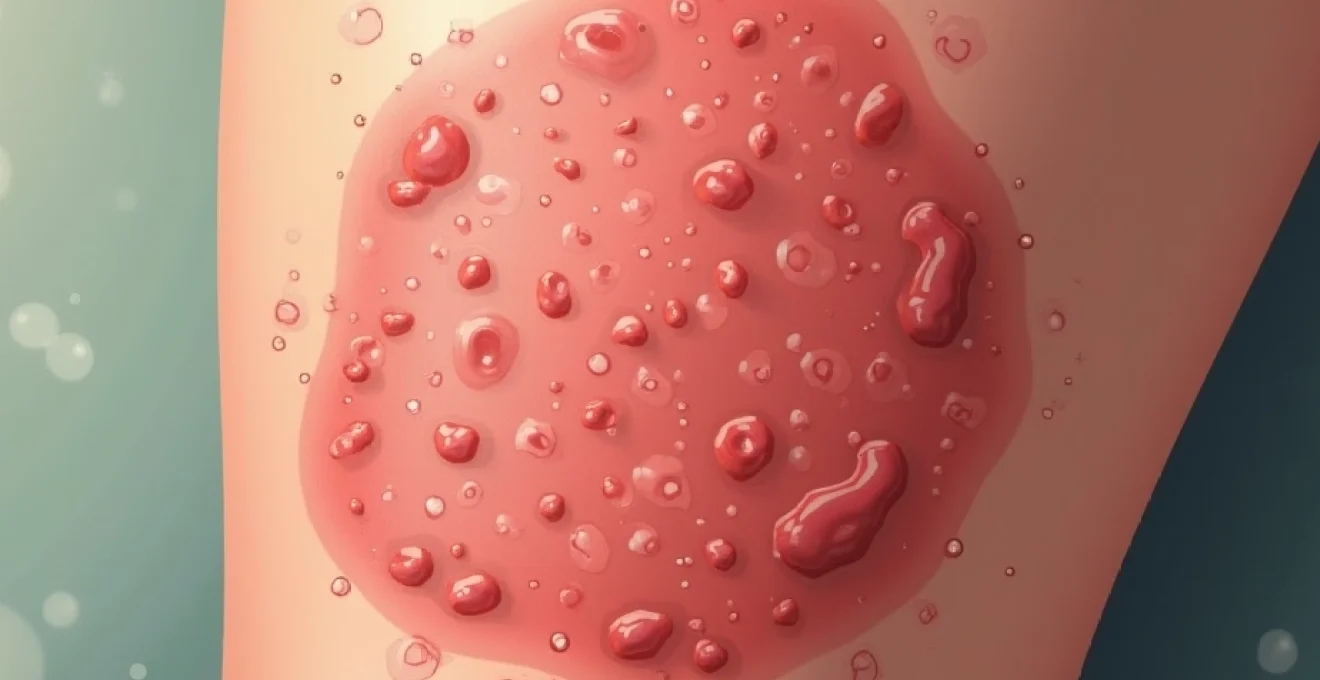
Cellulitis and bacterial skin infections: streptococcus and staphylococcus presentations
Cellulitis and bacterial skin infections can occasionally present with circular or ring-like patterns, particularly when the infection spreads centrifugally from a central point of inoculation. These serious bacterial infections primarily involve Streptococcus pyogenes and Staphylococcus aureus , with each organism demonstrating characteristic clinical presentations and progression patterns. Unlike superficial fungal infections, bacterial cellulitis extends into deeper dermal and subcutaneous tissues, creating more pronounced inflammatory responses including warmth, tenderness, and systemic symptoms.
The pathogenic mechanisms underlying circular bacterial infections involve initial breach of the skin barrier followed by rapid bacterial proliferation and toxin production. Streptococcus pyogenes produces hyaluronidase and streptokinase enzymes that facilitate tissue penetration and bacterial spread, whilst Staphylococcus aureus utilises various virulence factors including protein A and coagulase to establish infection. The circular expansion pattern may result from uniform bacterial dissemination outward from the initial inoculation site, though this presentation remains less common than the typical ill-defined borders characteristic of most cellulitis cases.
Bacterial cellulitis requires immediate antibiotic treatment to prevent serious complications including sepsis, necrotising fasciitis, and systemic spread of infection.
The clinical differentiation between circular cellulitis and other ring-shaped lesions relies on recognition of inflammatory signs including erythema, warmth, swelling, and tenderness. Cellulitis typically produces more pronounced systemic symptoms such as fever, malaise, and lymphangitic streaking, distinguishing it from superficial conditions like fungal infections or contact dermatitis. The presence of purulent drainage, fluctuance, or abscess formation suggests staphylococcal involvement, whilst rapidly spreading erythema with sharp, raised borders may indicate streptococcal cellulitis or erysipelas.
Treatment protocols for bacterial skin infections depend on severity assessment and likely causative organisms, with empirical antibiotic therapy targeting both streptococcal and staphylococcal species. Oral antibiotics such as flucloxacillin or erythromycin prove effective for mild cases, whilst severe infections require intravenous therapy with agents like flucloxacillin or vancomycin. The duration of treatment typically ranges from 7-14 days, with clinical monitoring essential to ensure adequate response and prevent complications. Failure to improve within 48-72 hours of appropriate antibiotic therapy warrants reassessment and consideration of alternative diagnoses or resistant organisms.
Autoimmune dermatological conditions: granuloma annulare and erythema annulare centrifugum
Autoimmune dermatological conditions including granuloma annulare and erythema annulare centrifugum represent important causes of circular rashes that require differentiation from infectious and allergic aetiologies. These conditions involve complex immunological processes targeting dermal components, resulting in characteristic inflammatory patterns that create ring-shaped lesions. Granuloma annulare demonstrates a predilection for dorsal surfaces of hands and feet but may affect any body area including the legs, whilst erythema annulare centrifugum typically presents with migratory annular plaques that expand peripherally over time
. These conditions present diagnostic challenges due to their non-infectious nature and potential association with underlying systemic diseases.
Granuloma annulare demonstrates characteristic histopathological features including central collagen degeneration surrounded by epithelioid cells and lymphocytes, creating the distinctive granulomatous inflammation pattern. The condition may occur in localised or generalised forms, with the localised variant more commonly affecting children and young adults whilst the generalised form typically presents in older individuals. Association with diabetes mellitus has been reported in some patients with widespread granuloma annulare, though the exact pathogenic relationship remains unclear.
Erythema annulare centrifugum presents as slowly expanding annular plaques with characteristic trailing scale on the inner aspect of the advancing border. This condition may be triggered by various factors including medications, infections, or malignancies, though many cases remain idiopathic. The migratory nature of these lesions distinguishes them from static conditions like tinea corporis, as the rings typically expand at rates of several millimetres per week whilst maintaining their distinctive morphology.
Histopathological analysis and dermal mucin deposits
Histopathological analysis of granuloma annulare reveals characteristic features that aid in definitive diagnosis when clinical appearance remains ambiguous. The dermal mucin deposits represent a hallmark finding, consisting of increased hyaluronic acid accumulation within the reticular dermis. These deposits stain positively with alcian blue and colloidal iron stains, creating the distinctive blue-grey appearance that helps differentiate granuloma annulare from other granulomatous conditions.
The inflammatory infiltrate in granuloma annulare demonstrates a characteristic pattern of lymphocytes and epithelioid cells arranged around areas of altered collagen. Central necrobiosis with palisading granulomatous inflammation creates the diagnostic histological appearance, though variations may occur depending on the lesion’s age and anatomical location. The absence of organisms on special stains helps exclude infectious causes, whilst the specific pattern of collagen alteration distinguishes granuloma annulare from other necrobiotic conditions.
Dermal mucin analysis provides additional diagnostic confirmation through quantitative assessment of glycosaminoglycan deposition. The increased mucin content reflects altered dermal matrix metabolism, possibly related to immune-mediated processes affecting collagen synthesis and degradation. This pathophysiological mechanism explains the clinical appearance of raised, firm papules that coalesce to form the characteristic annular configuration observed in active lesions.
Immunofluorescence patterns in autoimmune skin manifestations
Immunofluorescence patterns in granuloma annulare typically demonstrate negative direct immunofluorescence results, helping distinguish this condition from other autoimmune bullous diseases. However, indirect immunofluorescence may occasionally reveal circulating antibodies against dermal components, though these findings lack diagnostic specificity. The absence of characteristic immunofluorescence patterns supports the classification of granuloma annulare as a reactive rather than truly autoimmune condition.
Erythema annulare centrifugum demonstrates variable immunofluorescence findings depending on the underlying trigger and disease activity. Some cases may show deposition of immunoglobulins and complement components at the dermoepidermal junction, particularly when associated with underlying systemic conditions. The presence of perivascular immunoglobulin deposition may suggest immune complex-mediated pathogenesis, though this finding remains inconsistent across different patient populations.
Advanced immunohistochemical analysis reveals increased expression of inflammatory cytokines including tumour necrosis factor-alpha and interleukin-1 within the dermal infiltrate of both conditions. These findings support the role of inflammatory mediators in lesion development and progression, providing potential therapeutic targets for resistant cases. The cytokine profile may also help predict treatment response and guide selection of appropriate immunosuppressive therapies.
Corticosteroid treatment protocols and immunosuppressive therapies
Corticosteroid treatment protocols for granuloma annulare typically begin with high-potency topical preparations applied twice daily to affected areas. Intralesional corticosteroid injection represents an effective alternative for localised lesions, using triamcinolone acetonide 5-10mg/ml injected directly into the dermal component of active lesions. This approach provides higher local concentrations whilst minimising systemic exposure, making it particularly suitable for limited disease extent.
Systemic corticosteroid therapy may be considered for extensive or rapidly progressive cases, though the risk-benefit ratio requires careful evaluation given the benign nature of granuloma annulare. Prednisolone 0.5-1mg/kg daily for 4-6 weeks followed by gradual tapering may induce remission in severe cases, though recurrence rates remain high following discontinuation. Alternative immunosuppressive agents including methotrexate, ciclosporin, and hydroxychloroquine have shown efficacy in case reports and small series.
Immunosuppressive therapies for treatment-resistant autoimmune dermatological conditions require careful patient selection and monitoring protocols. Methotrexate therapy at doses of 10-25mg weekly demonstrates particular efficacy for generalised granuloma annulare, with response rates exceeding 60% in published series. Regular monitoring includes complete blood count, liver function tests, and chest radiography to detect potential adverse effects. The addition of folic acid supplementation reduces the risk of methotrexate-related toxicity whilst maintaining therapeutic efficacy.
Diagnostic methodology: dermatoscopy, skin biopsy, and laboratory analysis
Diagnostic methodology for circular rashes on the leg requires a systematic approach incorporating clinical examination, dermatoscopy, and selective use of laboratory investigations. Dermatoscopy provides valuable morphological details that aid in differential diagnosis, particularly for distinguishing between fungal infections and inflammatory conditions. The identification of specific dermatoscopic features including scale distribution, vascular patterns, and pigmentary changes helps narrow the diagnostic possibilities before proceeding to more invasive investigations.
Skin biopsy represents the gold standard for definitive diagnosis when clinical and dermatoscopic findings remain inconclusive. The selection of appropriate biopsy technique depends on lesion characteristics, with punch biopsy preferred for most circular lesions and incisional biopsy reserved for larger lesions requiring architectural assessment. Proper specimen handling including formalin fixation for routine histology and fresh tissue preservation for microbiological culture ensures optimal diagnostic yield.
Laboratory analysis encompasses multiple modalities tailored to suspected diagnoses, including direct microscopy, bacterial and fungal cultures, and serological testing. The integration of clinical findings with laboratory results provides the highest diagnostic accuracy whilst minimising unnecessary testing and associated costs. Modern molecular diagnostic techniques including polymerase chain reaction testing offer enhanced sensitivity for detecting specific pathogens, particularly in cases where traditional culture methods prove unsuccessful.
Accurate diagnosis of circular rashes requires correlation of clinical presentation, dermatoscopic findings, and appropriate laboratory investigations to ensure optimal patient outcomes and prevent complications.
The systematic approach to evaluating circular rashes begins with careful history-taking to identify potential exposures, travel history, medication use, and associated symptoms. Physical examination should assess lesion morphology, distribution, and associated findings such as lymphadenopathy or systemic signs of infection. Dermatoscopic examination provides additional morphological details that may not be apparent to naked-eye inspection, including subtle scale patterns, vascular architecture, and pigmentary variations that aid in differential diagnosis.
When definitive diagnosis remains uncertain following clinical assessment, tissue sampling through skin biopsy provides histopathological confirmation of suspected conditions. The selection of biopsy site should target the most active portion of the lesion, typically the advancing border for suspected infectious causes or the most inflamed area for suspected autoimmune conditions. Proper communication with the pathologist regarding clinical differential diagnosis ensures appropriate staining techniques and focused histopathological analysis.
Laboratory analysis protocols should be tailored to clinical suspicion, with KOH preparation and fungal culture indicated for suspected dermatophyte infections, bacterial culture for suspected cellulitis, and specialised testing for conditions like Lyme disease when epidemiologically appropriate. The judicious use of laboratory investigations based on clinical probability helps optimise diagnostic accuracy whilst controlling healthcare costs and minimising patient discomfort. Regular reassessment of diagnostic hypotheses ensures appropriate modification of testing strategies when initial results prove inconclusive or unexpected.