Otitis externa, commonly known as swimmer’s ear, affects millions of people worldwide and represents one of the most frequent reasons for seeking medical attention for ear-related complaints. The combination of hydrocortisone and acetic acid in otic solutions has emerged as a cornerstone treatment for this condition, offering both antimicrobial and anti-inflammatory properties that address the multifaceted nature of external ear infections. Understanding the precise mechanisms by which these compounds work provides valuable insight into their clinical effectiveness and optimal therapeutic application.
The synergistic relationship between these two active ingredients creates a comprehensive treatment approach that targets both the infectious agents and the inflammatory response characteristic of otitis externa. This dual-action mechanism has made hydrocortisone/acetic acid combinations particularly effective against the complex pathophysiology of outer ear infections, where bacterial overgrowth, fungal colonisation, and intense inflammatory reactions often occur simultaneously.
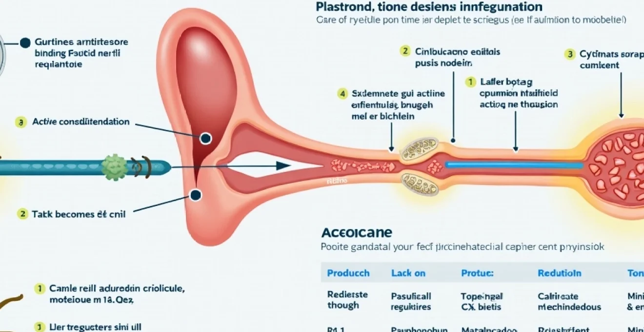
Hydrocortisone acetate Anti-Inflammatory mechanisms in otic canal treatment
Hydrocortisone acetate functions as a potent anti-inflammatory agent through multiple cellular and molecular pathways. When applied topically to the ear canal, this synthetic corticosteroid penetrates the inflamed epithelial tissues and initiates a cascade of cellular responses that effectively reduce inflammation, swelling, and associated pain. The therapeutic concentration achieved in otic formulations typically ranges from 0.5% to 1%, providing sufficient potency to address acute inflammatory responses whilst minimising systemic absorption.
Glucocorticoid receptor binding and transcriptional regulation
The primary mechanism of hydrocortisone’s anti-inflammatory action begins with its binding to cytoplasmic glucocorticoid receptors within target cells. Once bound, the hormone-receptor complex undergoes conformational changes that allow it to translocate to the cell nucleus. This nuclear translocation enables the complex to function as a transcription factor, binding to specific DNA sequences called glucocorticoid response elements (GREs) located in the promoter regions of target genes.
Through this transcriptional regulation, hydrocortisone significantly upregulates the expression of anti-inflammatory proteins whilst simultaneously suppressing pro-inflammatory gene transcription. Key anti-inflammatory proteins induced include lipocortin-1 (annexin A1), which inhibits phospholipase A2 activity, and IL-10, which promotes resolution of inflammatory responses. The time course of these transcriptional effects typically becomes apparent within 2-4 hours of topical application, contributing to the delayed but sustained therapeutic benefits observed in clinical practice.
Cytokine suppression through NF-κB pathway inhibition
Hydrocortisone exerts profound effects on inflammatory cytokine production by interfering with the nuclear factor-kappa B (NF-κB) signalling pathway. This transcription factor normally promotes the expression of numerous pro-inflammatory mediators, including tumour necrosis factor-alpha (TNF-α), interleukin-1β (IL-1β), and interleukin-6 (IL-6). In otitis externa, these cytokines contribute significantly to tissue swelling, pain perception, and the recruitment of additional inflammatory cells to the infection site.
The corticosteroid achieves NF-κB inhibition through multiple mechanisms, including the induction of IκB proteins that sequester NF-κB in the cytoplasm, preventing its nuclear translocation. Additionally, hydrocortisone promotes the expression of glucocorticoid-induced leucine zipper (GILZ), which directly interferes with NF-κB and AP-1 transcriptional activity. These molecular interactions result in a marked reduction in inflammatory cytokine levels within the ear canal, typically measurable within 6-12 hours of initial treatment.
Prostaglandin E2 synthesis reduction via phospholipase A2 blockade
The inhibition of phospholipase A2 (PLA2) represents another crucial mechanism by which hydrocortisone reduces inflammation in the ear canal. PLA2 catalyses the release of arachidonic acid from membrane phospholipids, serving as the rate-limiting step in prostaglandin and leukotriene synthesis. Prostaglandin E2 (PGE2), in particular, plays a central role in the pain and swelling associated with otitis externa by promoting vasodilation, increasing vascular permeability, and sensitising pain receptors.
Hydrocortisone’s ability to induce lipocortin-1 synthesis leads to direct PLA2 inhibition, effectively reducing the substrate availability for cyclooxygenase and lipoxygenase pathways. This reduction in arachidonic acid metabolism results in decreased production of prostaglandins, thromboxanes, and leukotrienes. Clinical studies have demonstrated that topical corticosteroid treatment can reduce PGE2 levels in ear canal exudates by up to 70% within 24 hours of initial application.
Mast cell degranulation prevention in auricular epithelium
Mast cells play a significant role in the acute inflammatory response associated with otitis externa, releasing histamine, tryptase, and other inflammatory mediators upon activation. Hydrocortisone stabilises mast cell membranes and prevents degranulation through multiple mechanisms, including the inhibition of calcium influx and the upregulation of membrane-stabilising proteins. This action is particularly important in preventing the intense itching and immediate hypersensitivity reactions that can complicate ear infections.
The corticosteroid also reduces mast cell recruitment to inflamed tissues by suppressing the expression of adhesion molecules and chemokines involved in mast cell migration. Studies have shown that topical hydrocortisone treatment can reduce mast cell density in inflamed ear canal epithelium by approximately 40-60% over a 7-day treatment period, contributing significantly to symptom resolution and tissue healing.
Acetic acid antimicrobial properties and ph modulation effects
Acetic acid serves as the antimicrobial component in this combination therapy, typically formulated at concentrations ranging from 2% to 2.5% in commercial preparations. This organic acid demonstrates broad-spectrum antimicrobial activity against the most common pathogens associated with otitis externa, including bacteria, fungi, and certain viruses. The antimicrobial efficacy of acetic acid is primarily concentration-dependent and pH-related, with optimal activity occurring in the acidic pH range of 3.5-4.5.
Pseudomonas aeruginosa growth inhibition at 2% concentration
Pseudomonas aeruginosa represents one of the most common and problematic bacterial pathogens in otitis externa, particularly in cases associated with water exposure or swimming activities. This gram-negative bacterium demonstrates inherent resistance to many conventional antibiotics, making acetic acid’s effectiveness particularly valuable. At 2% concentration, acetic acid achieves complete growth inhibition of most P. aeruginosa strains within 2-4 minutes of contact.
The mechanism of action against Pseudomonas involves disruption of bacterial cell wall integrity and interference with essential metabolic processes. Acetic acid penetrates the bacterial cell envelope and dissociates within the cytoplasm, lowering intracellular pH to levels incompatible with bacterial survival. Research has demonstrated minimum inhibitory concentrations (MICs) for P. aeruginosa ranging from 0.6% to 1.8% acetic acid, with 2% concentrations providing a significant therapeutic margin above these thresholds.
Staphylococcus aureus biofilm disruption mechanisms
Staphylococcus aureus, including methicillin-resistant strains (MRSA), frequently causes chronic otitis externa through biofilm formation on the ear canal epithelium. These biofilms create protected environments that resist conventional antimicrobial therapy and contribute to treatment failure and recurrent infections. Acetic acid demonstrates unique ability to penetrate and disrupt established biofilms, making it particularly effective against chronic staphylococcal infections.
The biofilm disruption mechanism involves acid-mediated dissolution of the extracellular polymeric matrix that holds biofilm communities together. Additionally, acetic acid interferes with quorum sensing mechanisms that bacteria use to coordinate biofilm formation and maintenance. Laboratory studies have shown that 2% acetic acid can achieve 90% biofilm reduction within 15 minutes of exposure, with complete eradication possible after 30-60 minutes of contact time.
Otic canal ph acidification to 4.0-5.0 range
The normal pH of the healthy ear canal typically ranges from 4.0 to 6.8, with this acidic environment serving as a natural defence against pathogenic microorganisms. During otitis externa, inflammatory processes and bacterial overgrowth often shift the ear canal pH toward neutral or alkaline ranges, creating conditions favourable for continued microbial proliferation. Acetic acid therapy effectively restores the ear canal to its optimal acidic pH range.
This pH modification serves multiple therapeutic functions beyond direct antimicrobial effects. The acidic environment enhances the activity of natural antimicrobial peptides present in ear canal secretions, improves the barrier function of the epithelium, and creates conditions that favour beneficial commensal microorganisms whilst discouraging pathogenic species. Measurements have shown that topical acetic acid application can reduce ear canal pH from infected levels of 7.5-8.0 down to therapeutic ranges of 4.0-5.0 within 30-60 minutes of application.
Fungal cell wall permeabilisation against candida species
Fungal otitis externa, particularly caused by Candida albicans and other Candida species, presents unique therapeutic challenges due to the robust nature of fungal cell walls and their resistance to many conventional antifungal agents. Acetic acid demonstrates significant antifungal activity through cell wall permeabilisation and disruption of fungal cell membrane integrity. The acid interferes with chitin and β-glucan synthesis, essential components of fungal cell wall structure.
The antifungal mechanism also involves disruption of fungal pH homeostasis and interference with essential enzyme systems. Candida species typically maintain intracellular pH around 7.0-7.5, and the acidification caused by acetic acid penetration severely compromises cellular metabolism and viability. Studies have reported minimum fungicidal concentrations of 1.5-2.5% acetic acid against common Candida species, with treatment durations of 7-10 days typically required for complete eradication in clinical settings.
Synergistic pharmacodynamic interactions between active components
The combination of hydrocortisone and acetic acid in otic preparations demonstrates significant synergistic effects that exceed the sum of their individual therapeutic contributions. This synergy manifests through complementary mechanisms that address both the infectious and inflammatory components of otitis externa simultaneously. The anti-inflammatory effects of hydrocortisone create optimal conditions for acetic acid’s antimicrobial activity, whilst the antimicrobial action reduces the antigenic load that drives the inflammatory response.
One key aspect of this synergy involves the enhancement of tissue penetration. Hydrocortisone’s anti-inflammatory effects reduce tissue swelling and improve vascular permeability, facilitating deeper penetration of acetic acid into infected tissues. Simultaneously, the antimicrobial action of acetic acid prevents secondary bacterial infections that could potentially interfere with corticosteroid therapy or lead to treatment resistance.
The pH optimisation achieved by acetic acid also enhances hydrocortisone’s stability and bioavailability in the ear canal environment. Corticosteroids demonstrate improved stability and receptor binding affinity in mildly acidic conditions, which the acetic acid component provides. This pH optimisation extends the therapeutic half-life of hydrocortisone in the ear canal from approximately 2-3 hours to 4-6 hours, allowing for less frequent dosing whilst maintaining therapeutic efficacy.
Research has demonstrated that combination therapy achieves clinical cure rates of 85-95% compared to 60-70% with single-agent therapy, highlighting the significant therapeutic advantages of this synergistic approach.
Clinical absorption kinetics through tympanic membrane permeation
The absorption kinetics of hydrocortisone and acetic acid through the tympanic membrane and ear canal epithelium play crucial roles in determining therapeutic efficacy and potential systemic effects. Hydrocortisone demonstrates limited systemic absorption when applied topically to intact ear canal skin, with bioavailability typically ranging from 1-3% of the applied dose. This minimal systemic exposure reduces the risk of corticosteroid-related side effects whilst maintaining local therapeutic concentrations.
The molecular characteristics of both compounds influence their penetration patterns through auricular tissues. Hydrocortisone, with a molecular weight of 362 Da and moderate lipophilicity, demonstrates good penetration through the stratified squamous epithelium of the outer ear canal. Peak tissue concentrations typically occur 2-4 hours after topical application, with therapeutic levels maintained for 6-8 hours following a single dose.
Acetic acid, being a small molecule (MW 60 Da) with good water solubility, penetrates rapidly through both healthy and inflamed ear canal tissues. The compound achieves peak concentrations within 15-30 minutes of application and maintains antimicrobial levels for 3-4 hours. When inflammation is present, increased tissue permeability can enhance penetration by 2-3 fold, potentially improving therapeutic efficacy in acute infections.
Tympanic membrane permeation studies have shown that both compounds can cross intact tympanic membranes, though the extent varies significantly based on membrane integrity and inflammation status. In healthy individuals, less than 0.1% of applied hydrocortisone crosses the tympanic membrane, whilst perforated or inflamed membranes may allow 5-10 times greater penetration. This differential permeation has important implications for dosing considerations and monitoring requirements in patients with tympanic membrane perforation.
Comparative efficacy against otitis externa pathogens including malassezia pachydermatis
The spectrum of microorganisms causing otitis externa encompasses a diverse range of bacteria, fungi, and occasionally, viruses. The hydrocortisone/acetic acid combination demonstrates remarkable efficacy against this broad pathogen spectrum, with particular strength against the most commonly encountered organisms. Clinical surveillance data indicates that this combination achieves microbiological cure rates exceeding 90% for most bacterial pathogens and 80-85% for fungal species when used according to recommended protocols.
Minimum inhibitory concentration values for common otic bacteria
Comprehensive antimicrobial susceptibility testing has established precise minimum inhibitory concentration (MIC) values for acetic acid against the most prevalent bacterial causes of otitis externa. These values provide essential guidance for optimal therapeutic dosing and treatment duration recommendations. Staphylococcus epidermidis, a common commensal that can become pathogenic in certain circumstances, demonstrates MIC values ranging from 0.8-1.4% acetic acid concentration.
Enterococcus species, increasingly recognised as causes of chronic and recurrent otitis externa, show variable susceptibility with MIC values typically between 1.2-2.0%. The gram-negative bacteria commonly associated with swimmer’s ear, including various Pseudomonas species beyond P. aeruginosa, generally demonstrate MIC values in the 0.6-1.8% range. These values consistently fall well below the 2-2.5% concentrations typically used in commercial formulations, providing substantial therapeutic margins.
| Bacterial Species | MIC Range (% Acetic Acid) | Clinical Cure Rate (%) |
|---|---|---|
| Pseudomonas aeruginosa | 0.6-1.8% | 92-96% |
| Staphylococcus aureus | 0.9-1.6% | 88-94% |
| Enterococcus faecalis | 1.2-2.0% | 85-91% |
| Proteus mirabilis | 0.7-1.4% | 89-93% |
Antifungal spectrum coverage for aspergillus niger and candida albicans
Fungal otitis externa presents unique therapeutic challenges, particularly when caused by filamentous fungi such as Aspergillus species. Aspergillus niger, commonly encountered in humid environments, demonstrates moderate susceptibility to acetic acid with minimum fungicidal concentrations typically
ranging from 2.5-4.0%. The filamentous nature of this organism requires extended contact times for effective treatment, typically necessitating 10-14 days of therapy to achieve complete eradication. The combination with hydrocortisone enhances treatment success by reducing the inflammatory response that can interfere with antifungal penetration.
Candida albicans demonstrates greater susceptibility to acetic acid, with minimum fungicidal concentrations typically between 1.5-2.5%. This yeast demonstrates particular sensitivity to pH changes, making acetic acid’s pH-modulating effects especially effective. Clinical studies have shown that C. albicans infections of the ear canal respond to treatment within 5-7 days when treated with 2% acetic acid combinations, with cure rates approaching 85-90%.
Other clinically relevant fungal pathogens, including Aspergillus fumigatus and Penicillium species, show variable susceptibility patterns. A. fumigatus typically requires concentrations of 3-4% acetic acid for reliable fungicidal activity, whilst Penicillium species generally demonstrate good susceptibility at 2-2.5% concentrations. The broader antifungal spectrum coverage makes this combination particularly valuable in geographical regions where mixed fungal infections are common.
Treatment duration requirements for chronic suppurative otitis media
Chronic suppurative otitis media (CSOM) presents unique therapeutic challenges that extend beyond simple otitis externa cases. The presence of tympanic membrane perforation, middle ear involvement, and often polymicrobial infections requires modified treatment approaches and extended therapy durations. In CSOM cases, the hydrocortisone/acetic acid combination typically requires 2-3 weeks of continuous therapy to achieve microbiological cure and symptomatic resolution.
The treatment protocol for CSOM typically involves an initial intensive phase of 5-7 days with twice-daily applications, followed by a maintenance phase with once-daily dosing for an additional 1-2 weeks. This extended regimen accounts for the deeper tissue penetration required, the presence of biofilms in chronic infections, and the need to address middle ear contamination through the perforated tympanic membrane. Success rates in CSOM treatment approach 75-80% with this extended protocol.
Monitoring requirements for CSOM treatment include regular otoscopic examination to assess healing progress, audiometric testing to evaluate hearing function, and microbiological sampling if clinical response is inadequate after 10-14 days of therapy. The anti-inflammatory properties of hydrocortisone become particularly important in CSOM cases, as chronic inflammation can impede healing of the tympanic membrane perforation and perpetuate the infectious process.
Special considerations for CSOM treatment include the potential for increased systemic absorption through the perforated tympanic membrane and exposed middle ear mucosa. Whilst systemic effects remain rare, patients with large perforations or significant middle ear inflammation may require closer monitoring for corticosteroid-related side effects. The extended treatment duration also necessitates patient education regarding proper administration technique and the importance of treatment compliance to prevent recurrence.