Nerve repair surgery represents one of the most complex and challenging procedures in reconstructive microsurgery, requiring both surgical precision and extensive patient commitment during the recovery process. The journey from initial surgical intervention to functional restoration involves intricate biological mechanisms that unfold over months or even years. Understanding the timeline and factors that influence recovery becomes essential for patients facing this procedure, as realistic expectations can significantly impact both psychological wellbeing and compliance with rehabilitation protocols. The complexity of nerve regeneration means that recovery varies dramatically between individuals, with some experiencing meaningful improvements within six months whilst others may require two years or more to achieve optimal outcomes.
Modern surgical techniques have revolutionised nerve repair outcomes, yet the fundamental biological limitations of neural regeneration remain unchanged. The peripheral nervous system possesses remarkable regenerative capacity compared to the central nervous system, but this process occurs at a predictably slow pace that tests patient patience and determination. Surgeons now utilise advanced microsurgical techniques, sophisticated nerve grafting procedures, and innovative conduit technologies to optimise conditions for regeneration, yet the ultimate success depends heavily on the body’s innate healing mechanisms and the patient’s commitment to comprehensive rehabilitation.
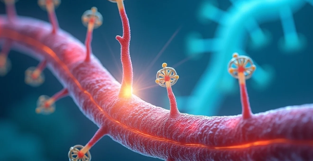
Peripheral nerve regeneration timeline following microsurgical repair
The biological process of nerve regeneration follows a predictable yet highly variable timeline that begins immediately after surgical repair and continues for months or years. Understanding this complex cascade of cellular events helps patients appreciate why nerve recovery cannot be rushed and why consistent rehabilitation remains crucial throughout the extended healing period. The regeneration process encompasses multiple overlapping phases, each with distinct cellular mechanisms and clinical implications for functional recovery.
Wallerian degeneration phase and initial cellular response
The initial phase following nerve repair involves Wallerian degeneration, a process where the distal nerve segment undergoes controlled breakdown and clearance. This phase typically begins within 24 to 48 hours post-surgery and continues for approximately four weeks. During this period, the myelin sheath surrounding damaged axons degenerates, and macrophages infiltrate the injury site to clear debris. Patients often experience complete loss of function in the affected area during this phase, which can be psychologically challenging but represents a necessary biological prerequisite for regeneration.
The surgical repair site requires approximately three to six weeks for the outer nerve sheath, called the epineurium, to heal sufficiently to provide structural support for regenerating axons. During this critical period, patients must wear protective splinting to prevent tension on the repair site that could disrupt healing. The cellular environment during Wallerian degeneration involves complex molecular signalling that either promotes or inhibits subsequent regeneration, making this phase crucial for long-term outcomes.
Axonal sprouting and growth cone formation mechanisms
Following the degeneration phase, surviving axons begin forming growth cones at their cut ends, initiating the sprouting process that will eventually restore neural connections. This phase typically commences around the fourth week post-surgery and represents the beginning of active regeneration. Multiple axonal sprouts emerge from each surviving nerve fibre, creating redundancy that improves the likelihood of successful target reinnervation. However, this redundancy also means that many sprouts will eventually be eliminated through a process called pruning.
The growth cone formation involves sophisticated molecular machinery that enables axons to navigate through the healing tissue environment. Growth factors, adhesion molecules, and guidance cues direct axonal growth towards appropriate targets, though this process remains imperfect even under optimal conditions. Patients may begin experiencing tingling sensations or electrical shooting pains during this phase, indicating that axonal sprouting has commenced and growth cones are beginning their journey towards target tissues.
Schwann cell proliferation and remyelination process
Concurrent with axonal sprouting, Schwann cells proliferate and form guidance tubes called Bands of Büngner that provide structural support for regenerating axons. These cells play a crucial role in nerve regeneration by secreting growth factors, clearing debris, and ultimately remyelinating successfully regenerated axons. The Schwann cell response begins within days of injury but reaches peak activity during the second and third months following repair.
The remyelination process occurs gradually as regenerating axons reach their targets and establish functional connections. This phase can extend from three months to over a year post-surgery, depending on the distance axons must travel and the complexity of target reinnervation. Successful remyelination dramatically improves nerve conduction velocity and functional outcomes, but the process remains incomplete in many cases, explaining why nerve function rarely returns to pre-injury levels.
The regeneration rate of peripheral nerves follows a remarkably consistent pattern of approximately one millimetre per day once active growth begins, making the distance from repair site to target tissue the primary determinant of recovery timeline.
Target organ reinnervation and functional recovery stages
The final phase of nerve regeneration involves target organ reinnervation, where regenerating axons must locate and innervate appropriate muscle fibres or sensory receptors. This process typically begins three to six months post-surgery for proximal repairs but may be delayed up to a year or more for repairs involving significant nerve gaps or distal locations. Motor reinnervation faces particular time constraints, as muscle fibres undergo irreversible changes if denervated for more than 18 to 24 months.
Functional recovery occurs gradually as newly formed neural connections mature and strengthen through use. Initial motor recovery often appears as weak, uncoordinated movements that gradually improve with rehabilitation. Sensory recovery typically follows a predictable pattern, beginning with crude touch and pressure sensation before progressing to more sophisticated discriminatory functions. The quality of functional recovery depends heavily on the accuracy of axonal pathfinding and the patient’s commitment to rehabilitation exercises that promote neural plasticity and strengthen new connections.
Factors influencing postoperative recovery duration in nerve reconstruction
Multiple interconnected factors influence the duration and extent of recovery following nerve repair surgery, creating significant variability in outcomes between patients with seemingly similar injuries. Understanding these variables helps both surgeons and patients establish realistic expectations and develop appropriate treatment strategies. The interplay between biological factors, surgical variables, and patient-specific characteristics creates a complex equation that determines ultimate functional outcomes.
Patient age and neuroplasticity considerations
Age represents one of the most significant factors influencing nerve regeneration capacity, with younger patients demonstrating superior regenerative potential and functional recovery compared to older individuals. Children and young adults benefit from enhanced neuroplasticity, allowing their nervous systems to adapt more effectively to altered innervation patterns and compensate for incomplete regeneration. The regenerative capacity of peripheral nerves declines progressively with age, partly due to reduced growth factor production and decreased Schwann cell responsiveness.
Neuroplasticity considerations extend beyond simple regeneration to encompass the brain’s ability to reorganise and adapt to changed sensory input patterns. Younger patients more readily adapt to altered sensation patterns and can often achieve functional outcomes that exceed what would be predicted based purely on anatomical regeneration. Adult patients may require more intensive sensory reeducation programmes to maximise their functional recovery potential, whilst elderly patients may face additional challenges related to concurrent medical conditions that impair healing.
Nerve gap distance and surgical technique selection
The distance between nerve ends significantly influences both the surgical approach and recovery timeline, with larger gaps requiring more complex reconstruction techniques and longer regeneration periods. Direct nerve repair, possible when minimal tissue loss has occurred, offers the best potential for recovery with shorter healing times. However, when nerve gaps exceed 2-3 centimetres, surgeons must utilise nerve grafts or synthetic conduits, which introduce additional variables and typically result in less predictable outcomes.
Nerve grafting involves harvesting sensory nerves from other body areas to bridge the gap between injured nerve ends, creating multiple repair sites that regenerating axons must navigate successfully. This technique, whilst effective for bridging larger defects, typically results in some degree of donor site morbidity and may achieve only 60-80% of normal function even in successful cases. Synthetic nerve conduits represent an evolving technology that eliminates donor site morbidity but currently show inferior results compared to autologous nerve grafts for gaps exceeding 3 centimetres.
Location-specific recovery variables in upper and lower extremities
The anatomical location of nerve injury significantly influences recovery patterns, with proximal injuries generally requiring longer regeneration periods but potentially achieving better functional outcomes due to the presence of larger nerve fascicles and more robust regenerative capacity. Upper extremity nerve injuries often receive priority attention due to their impact on hand function and occupational capacity, whilst lower extremity injuries may focus more on protective sensation and basic motor function.
Digital nerve repairs in the fingers represent relatively favourable injuries due to the short regeneration distance and specific functional requirements. These repairs often demonstrate meaningful recovery within 6-9 months, though complete restoration of two-point discrimination may require up to two years. Conversely, brachial plexus injuries involving nerve roots may require multiple procedures and years of rehabilitation, with recovery outcomes varying dramatically based on the specific nerves involved and the mechanism of injury.
Comorbidities impact on nerve healing capacity
Systemic health conditions significantly influence nerve regeneration capacity, with diabetes mellitus representing the most common and problematic comorbidity affecting nerve healing. Diabetic patients experience delayed wound healing, reduced growth factor production, and compromised Schwann cell function that collectively impair regeneration outcomes. Smoking represents another major modifiable risk factor, as nicotine and other toxins reduce tissue oxygenation and impair cellular metabolism essential for nerve regeneration.
Nutritional status plays a crucial but often overlooked role in nerve regeneration, with deficiencies in vitamins B12, B6, and folate potentially compromising recovery outcomes. Chronic inflammatory conditions, autoimmune disorders, and certain medications may also negatively impact nerve healing capacity. Patients with multiple comorbidities may require extended recovery periods and more intensive rehabilitation programmes to achieve optimal outcomes, emphasising the importance of comprehensive medical optimisation before and after nerve repair surgery.
Surgical Technique-Specific recovery expectations
Different surgical approaches to nerve repair carry distinct recovery profiles and outcome expectations that patients must understand when making treatment decisions. Primary nerve repair, performed immediately after injury when conditions permit, typically offers the most favourable prognosis with recovery beginning within 3-4 months and potentially achieving 70-90% of normal function in ideal cases. The direct coaptation of nerve ends minimises the regeneration distance and provides optimal conditions for axonal growth, though successful outcomes still require months of patient rehabilitation and realistic expectation management.
Secondary nerve reconstruction, performed weeks or months after initial injury, faces additional challenges related to scar tissue formation and muscle atrophy that occurred during the delay period. These procedures often require more complex surgical techniques, including neurolysis to free nerves from scar tissue and tendon transfers to restore function in cases where nerve regeneration may be incomplete. Recovery timelines extend significantly, often requiring 12-24 months before meaningful function returns, and ultimate outcomes may be more limited compared to primary repairs.
Nerve grafting procedures introduce multiple variables that influence recovery patterns, as regenerating axons must successfully navigate through both the proximal and distal repair sites. The number and length of grafts required correlates with recovery complexity, with longer grafts and multiple grafts generally associated with more prolonged and less predictable recovery courses. Successful nerve graft procedures may achieve 60-80% of normal function, but patients must understand that complete restoration rarely occurs and that recovery may continue for two years or longer.
Modern microsurgical techniques have transformed nerve repair outcomes, yet the biological limitations of neural regeneration mean that even expertly performed procedures require extensive time and patient commitment to achieve optimal results.
Synthetic nerve conduits and processed nerve allografts represent emerging technologies that may eliminate the need for autologous nerve harvesting whilst providing structural guidance for regenerating axons. Early clinical results suggest these materials may be effective for gaps up to 3-4 centimetres, though long-term outcomes remain under investigation. Patients receiving these newer technologies should understand that they represent evolving treatments with less established outcome data compared to traditional nerve grafting techniques, though they offer the advantage of eliminating donor site morbidity.
Postoperative rehabilitation protocols and recovery milestones
Comprehensive rehabilitation following nerve repair surgery encompasses multiple phases that correspond to the biological stages of nerve regeneration, with each phase requiring specific interventions and patient education to optimise outcomes. The immediate postoperative period focuses on wound healing and protection of the repair site through appropriate splinting and activity modification. Patients typically wear protective splints for 3-6 weeks, during which time gentle range of motion exercises for uninvolved joints help prevent secondary complications whilst protecting the healing nerve repair.
The early mobilisation phase begins once adequate healing has occurred at the repair site, typically 4-6 weeks post-surgery, and focuses on maintaining joint mobility and preventing contractures in denervated areas. Passive range of motion exercises become crucial during this phase, as patients cannot actively control affected muscles but must prevent joint stiffness that could compromise functional outcomes once reinnervation begins. Sensory reeducation programmes commence early, even before sensation returns, to maintain cortical representation of the affected body part and facilitate integration of returning sensation.
The intermediate rehabilitation phase, occurring 3-6 months post-surgery, typically coincides with the earliest signs of motor and sensory recovery. Patients may notice tingling sensations, weak muscle contractions, or Tinel’s sign progression as regenerating axons advance towards their targets. Rehabilitation focus shifts towards strengthening exercises for recovering muscles, progressive sensory discrimination training, and functional task practice. This phase requires careful monitoring to adjust exercise intensity based on recovery progress whilst avoiding overexertion that could impede healing.
Advanced rehabilitation continues for months or years as functional recovery progresses, emphasising task-specific training, sensory integration, and compensation strategies for incomplete recovery. Patients learn to maximise the function of recovering nerves whilst developing alternative strategies for tasks that remain challenging. The rehabilitation team monitors progress through objective measures including strength testing, sensory mapping, and functional outcome assessments that guide treatment modifications and help establish realistic long-term goals.
Successful nerve repair rehabilitation requires a partnership between patient, surgeon, and therapist that extends far beyond the initial surgery, with consistent engagement over many months determining the ultimate functional outcome.
Complications and delayed recovery scenarios in nerve repair surgery
Despite optimal surgical technique and patient compliance, nerve repair procedures may encounter complications that delay or limit recovery outcomes, requiring additional interventions and modified expectations. Infection represents an immediate postoperative concern that can significantly impair nerve regeneration through inflammatory damage and scar tissue formation. Early recognition and aggressive treatment of infection become crucial for preserving regenerative potential, with some cases requiring surgical debridement and revision repair procedures that further delay recovery timelines.
Scar tissue formation at the repair site can impede axonal regeneration and create painful neuromas that require surgical intervention. Neuroma formation occurs when regenerating axons fail to find appropriate targets and instead form disorganised nerve tissue that can be exquisitely painful and functionally limiting. Surgical neuroma revision may be necessary months or years after the initial repair, essentially restarting the regeneration process and extending overall recovery time. Prevention through meticulous surgical technique and appropriate postoperative care remains the preferred approach to managing this complication.
Incomplete or misdirected regeneration represents a common outcome that may necessitate additional procedures such as tendon transfers or nerve transfers to restore functional capacity. When regenerating axons fail to reach appropriate targets or innervate incorrect structures, patients may experience limited recovery despite adequate regeneration time. These scenarios often require creative surgical solutions and intensive rehabilitation to maximise remaining function whilst accepting permanent limitations in some areas.
Complex regional pain syndrome occasionally develops following nerve repair surgery, creating chronic pain and functional limitations that may overshadow the benefits of successful nerve regeneration. This challenging condition requires multidisciplinary management approaches that may include medications, nerve blocks, psychological support, and intensive physical therapy. Early recognition and intervention provide the best outcomes for managing this complication, though some patients may experience long-term functional limitations that require ongoing management strategies and lifestyle modifications.