Abdominal lymph node inflammation represents a complex immunological response that affects millions of patients worldwide, ranging from benign infectious processes to serious malignant conditions. The intricate network of lymphatic structures within the abdomen serves as a crucial battleground where immune cells encounter pathogens, malignant cells, and inflammatory mediators. Understanding the mechanisms behind lymphadenopathy in abdominal compartments requires a comprehensive appreciation of anatomical relationships, pathophysiological processes, and clinical manifestations that distinguish various aetiologies.
The significance of abdominal lymphadenopathy extends beyond simple immune responses, as these nodes often serve as sentinels for systemic disease processes. From mesenteric adenitis in paediatric populations to retroperitoneal lymphadenopathy in malignant conditions, the clinical spectrum encompasses both acute self-limiting conditions and chronic progressive disorders that require immediate therapeutic intervention.
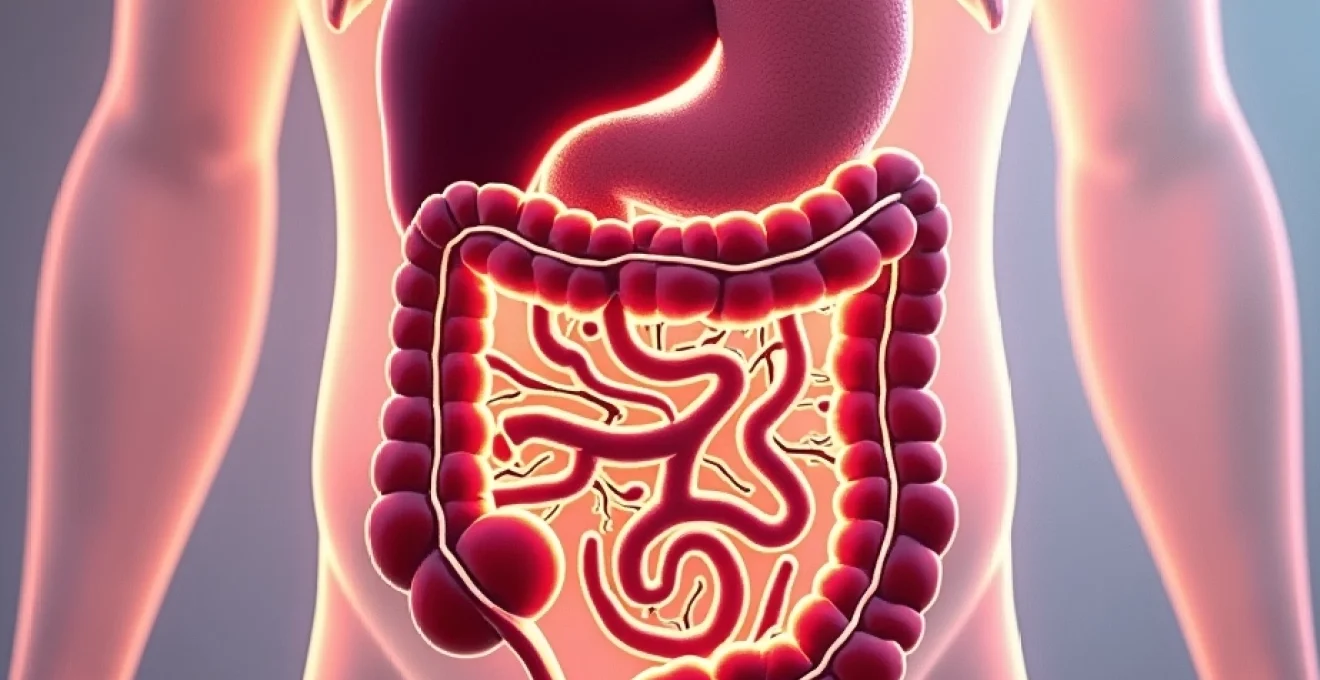
Anatomical distribution and classification of abdominal lymph nodes
The abdominal cavity contains an extensive network of lymphatic structures organised into distinct anatomical regions, each with specific drainage patterns and clinical significance. These lymph nodes are strategically positioned to monitor lymphatic fluid from various organs, creating a sophisticated surveillance system that can detect both infectious agents and malignant cells as they traverse the lymphatic circulation.
Mesenteric lymph node networks and drainage patterns
The mesenteric lymph nodes form the largest concentration of lymphoid tissue within the abdomen, with hundreds of nodes distributed throughout the small bowel and colonic mesentery. These nodes are organised into three distinct groups: the ileocolic nodes, which drain the terminal ileum and caecum; the right colic nodes, serving the ascending colon; and the middle colic nodes, responsible for transverse colon drainage. The superior mesenteric lymph node chain represents the central collection point for lymphatic drainage from the entire small bowel, while the inferior mesenteric nodes collect lymph from the left colon and upper rectum.
Mesenteric lymphadenopathy frequently manifests as mesenteric adenitis , particularly in children and adolescents, where viral infections commonly trigger inflammatory responses. The condition often presents with right lower quadrant pain that can mimic appendicitis, creating diagnostic challenges for clinicians. Understanding the normal size parameters—typically less than 5 millimetres in healthy individuals—becomes crucial when evaluating pathological enlargement during imaging studies.
Retroperitoneal lymphatic chains and para-aortic node groups
The retroperitoneal space houses several critical lymph node groups that drain deep abdominal structures and pelvic organs. The para-aortic lymph nodes extend from the diaphragm to the aortic bifurcation, forming left and right chains that receive lymphatic drainage from the kidneys, adrenal glands, gonads, and portions of the gastrointestinal tract. These nodes are particularly significant in oncological staging, as their involvement often indicates advanced disease in various malignancies.
The paracaval lymph nodes, situated along the inferior vena cava, work in conjunction with the para-aortic chains to create a comprehensive filtration system for retroperitoneal structures. Inflammatory conditions affecting these node groups often present with deep, poorly localised abdominal pain and may be associated with systemic symptoms such as fever and weight loss. The anatomical proximity to major vascular structures makes surgical intervention in this region particularly challenging.
Coeliac and superior mesenteric artery node classifications
The coeliac axis lymph nodes are strategically positioned around the coeliac trunk and its major branches, creating distinct subgroups based on their relationship to arterial structures. The left gastric nodes drain the lesser curvature of the stomach and lower oesophagus, while the hepatic artery nodes receive lymphatic drainage from the liver, gallbladder, and pancreatic head. The splenic artery nodes serve the spleen, pancreatic tail, and greater curvature of the stomach.
Superior mesenteric artery lymph nodes form a crucial junction point where lymphatic vessels from the small bowel converge before proceeding to the cisterna chyli. These nodes are frequently involved in inflammatory bowel disease, particularly Crohn’s disease, where chronic inflammation leads to characteristic creeping fat and lymph node enlargement. The superior mesenteric nodes also serve as important staging markers in pancreatic and small bowel malignancies.
Porta hepatis and hepatoduodenal ligament lymphatic structures
The porta hepatis represents a critical anatomical region where multiple lymph node groups converge around the hepatic artery, portal vein, and bile duct structures. These nodes, collectively known as the hepatoduodenal ligament lymph nodes, serve as primary drainage sites for the liver, gallbladder, pancreatic head, and duodenum. Their strategic location makes them essential markers for hepatobiliary and pancreatic pathology.
Portal lymphadenopathy often indicates serious underlying conditions, including cholangiocarcinoma, pancreatic adenocarcinoma, or metastatic disease from gastrointestinal primaries. The close anatomical relationship between these nodes and vital structures complicates both diagnostic and therapeutic approaches, requiring careful multidisciplinary planning when intervention becomes necessary.
Pathophysiology of lymphadenopathy in abdominal compartments
The development of lymph node inflammation within abdominal compartments involves complex interactions between immune cells, inflammatory mediators, and tissue architecture that result in characteristic morphological and functional changes. Understanding these pathophysiological mechanisms provides crucial insights into disease progression and therapeutic targets.
Cytokine-mediated inflammatory cascades in lymphoid tissue
Lymph node inflammation initiates through activation of resident antigen-presenting cells, primarily dendritic cells and macrophages, which encounter foreign antigens or damage-associated molecular patterns. These activated cells rapidly release pro-inflammatory cytokines including interleukin-1β, tumour necrosis factor-α, and interleukin-6, creating a local inflammatory environment that attracts circulating immune cells to the lymph node.
The cytokine cascade triggers vasodilation and increased vascular permeability within the lymph node architecture, leading to the characteristic swelling and tenderness associated with lymphadenopathy. Chemokines such as CCL19 and CCL21 facilitate T-cell trafficking to appropriate anatomical compartments within the node, while interferons enhance antigen presentation capabilities and promote effective immune responses against infectious agents.
Antigen presentation and T-Cell activation mechanisms
Effective lymph node function depends on sophisticated antigen presentation mechanisms that occur within specialised microenvironments. Dendritic cells capture antigens in peripheral tissues and migrate to regional lymph nodes, where they present processed peptides to naive T-cells within the T-cell zones. This process requires upregulation of co-stimulatory molecules and major histocompatibility complex expression, creating the immunological synapse necessary for T-cell activation.
B-cell responses occur simultaneously within germinal centres, where antigen-specific B-cells undergo somatic hypermutation and class switching under the guidance of follicular helper T-cells. The formation of germinal centres contributes significantly to lymph node enlargement and represents a hallmark of effective adaptive immune responses. Dysregulation of these processes can lead to persistent lymphadenopathy or inappropriate immune responses.
Complement system activation and immune complex formation
The complement system plays a crucial role in lymph node inflammation through multiple activation pathways that enhance immune responses and contribute to tissue changes. Classical pathway activation occurs when antibodies bind to antigens, creating immune complexes that activate C1q and initiate the complement cascade. Alternative pathway activation provides innate immune recognition of pathogen-associated molecular patterns without requiring antibody involvement.
Complement activation products, particularly C3a and C5a, serve as powerful chemoattractants that recruit neutrophils and monocytes to inflamed lymph nodes. The membrane attack complex formation can directly damage pathogen membranes while complement opsonisation enhances phagocytosis by macrophages and neutrophils within the lymph node architecture. Immune complex deposition can lead to chronic inflammation and structural changes within affected nodes.
Vascular permeability changes and lymphatic vessel dilation
Inflammatory mediators significantly alter vascular permeability within lymph nodes, allowing increased protein extravasation and cellular infiltration that contributes to node enlargement. Histamine, leukotrienes, and prostaglandins directly affect endothelial cell junctions, while nitric oxide and other vasoactive substances cause smooth muscle relaxation and vasodilation. These changes facilitate immune cell trafficking but can also contribute to tissue oedema and pain.
Lymphatic vessel dilation occurs as a compensatory mechanism to handle increased lymph flow during inflammatory responses. The smooth muscle cells within lymphatic vessel walls respond to inflammatory mediators by relaxing, increasing vessel calibre and reducing resistance to lymph flow. However, excessive dilation can impair the normal pumping function of lymphatic vessels, leading to lymphatic stasis and further node enlargement.
Infectious aetiologies and microbial patterns
Infectious causes represent the most common aetiology of abdominal lymph node inflammation, with distinct microbial patterns producing characteristic clinical presentations and imaging findings. Understanding these infectious patterns enables clinicians to develop targeted diagnostic and therapeutic approaches while recognising potential complications and long-term sequelae.
Mycobacterium tuberculosis and atypical mycobacterial infections
Mycobacterial infections produce distinctive lymphadenopathy characterised by granulomatous inflammation and caseous necrosis within affected nodes. Mycobacterium tuberculosis typically causes abdominal lymphadenopathy as part of disseminated disease or through direct extension from pulmonary infection. The organisms have a predilection for mesenteric and retroperitoneal lymph nodes, where they can remain dormant for extended periods before reactivating during immunocompromised states.
Atypical mycobacteria, particularly Mycobacterium avium complex , frequently cause abdominal lymphadenopathy in immunocompromised patients, especially those with HIV/AIDS. These infections often present with massive lymph node enlargement and systemic symptoms including fever, night sweats, and weight loss. The characteristic imaging appearance shows central necrosis with rim enhancement, though this finding is not pathognomonic for mycobacterial infection.
The granulomatous response to mycobacterial infection creates a unique histological pattern that helps distinguish tuberculous lymphadenitis from other infectious causes, though definitive diagnosis requires microbiological confirmation through culture or molecular methods.
Epstein-barr virus and Cytomegalovirus-Associated lymphadenitis
Viral infections, particularly those caused by Epstein-Barr virus (EBV) and cytomegalovirus (CMV), frequently trigger abdominal lymphadenopathy as part of systemic lymphoproliferative responses. EBV infection can cause massive splenomegaly and generalised lymphadenopathy, including involvement of mesenteric and retroperitoneal node groups. The virus preferentially infects B-lymphocytes, leading to polyclonal B-cell proliferation and characteristic atypical lymphocytes in peripheral blood.
CMV infections typically produce more subtle lymph node changes but can cause significant abdominal lymphadenopathy in immunocompromised patients or during primary infection in healthy individuals. The virus has tropism for multiple cell types, including lymphocytes, monocytes, and endothelial cells, creating diverse clinical presentations. Both viral infections can trigger hemophagocytic lymphohistiocytosis, a potentially fatal syndrome characterised by excessive immune activation and multi-organ dysfunction.
Yersinia enterocolitica and salmonella species involvement
Yersinia enterocolitica represents a particularly important cause of mesenteric lymphadenitis, especially in children and young adults, where it can closely mimic acute appendicitis. The organism preferentially invades Peyer’s patches and mesenteric lymph nodes, causing acute inflammation that predominantly affects the terminal ileum and right-sided colonic lymphatic drainage. Geographic variation in Y. enterocolitica serotypes influences clinical presentation and severity of lymph node involvement.
Salmonella species, including both typhoidal and non-typhoidal strains, can cause significant abdominal lymphadenopathy through different mechanisms. Salmonella typhi produces systemic infection with characteristic involvement of mesenteric lymph nodes, while non-typhoidal Salmonella typically causes more localised gastroenteritis with reactive lymphadenopathy. The organisms’ ability to survive within macrophages contributes to persistent infection and chronic lymph node enlargement in some patients.
Bartonella henselae and cat scratch disease manifestations
Bartonella henselae infection, commonly known as cat scratch disease, can produce significant abdominal lymphadenopathy, particularly affecting mesenteric and retroperitoneal node groups. The organism typically enters through skin inoculation following cat scratches or bites, then disseminates through lymphatic channels to regional and distant lymph nodes. Parinaud’s oculoglandular syndrome represents a characteristic presentation with conjunctival lesions and preauricular lymphadenopathy.
Atypical presentations of cat scratch disease can include isolated abdominal lymphadenopathy without obvious cutaneous lesions, making diagnosis challenging. The organism’s slow growth characteristics and fastidious culture requirements often necessitate serological or molecular diagnostic approaches. Treatment typically involves macrolide antibiotics, though many cases resolve spontaneously without antimicrobial intervention.
Neoplastic causes and malignant transformation patterns
Malignant conditions represent serious causes of abdominal lymphadenopathy that require prompt recognition and aggressive treatment. Primary lymphoid malignancies and metastatic disease from solid organ tumours create distinct patterns of lymph node involvement that influence staging, prognosis, and therapeutic approaches. Understanding these neoplastic processes enables clinicians to differentiate malignant from benign causes of lymphadenopathy and implement appropriate diagnostic strategies.
Primary lymphomas, including both Hodgkin and non-Hodgkin subtypes, frequently involve abdominal lymph node groups as part of their natural disease progression. Diffuse large B-cell lymphoma represents the most common aggressive lymphoma subtype, often presenting with rapidly enlarging abdominal masses and constitutional symptoms. The disease typically affects multiple node groups simultaneously and can involve extranodal sites including the gastrointestinal tract, liver, and spleen.
Hodgkin lymphoma demonstrates characteristic contiguous spread patterns that distinguish it from non-Hodgkin lymphomas, which tend to present with more random distribution of nodal involvement. The presence of Reed-Sternberg cells within an inflammatory background creates the pathognomonic histological appearance of Hodgkin lymphoma, though immunohistochemical studies are essential for definitive subtyping and treatment planning.
Metastatic lymphadenopathy results from haematogenous or lymphatic spread of solid organ malignancies to regional or distant lymph node groups. Common primary sites include gastric, pancreatic, and colorectal adenocarcinomas, which frequently metastasise to corresponding regional lymph node basins. The pattern of lymph node involvement often reflects the natural lymphatic drainage pathways from the primary tumour site, though skip lesions and retrograde spread can occur.
The distinction between reactive and malignant lymphadenopathy relies heavily on imaging characteristics, clinical context, and ultimately histopathological examination, as no single feature can reliably differentiate benign from malignant causes.
Gynaecological malignancies, particularly ovarian and endometrial cancers, commonly involve para-aortic and pelvic lymph node groups as part of their staging workup. The International Federation of Gynaecology and Obstetrics staging systems incorporate lymph node involvement as a critical prognostic factor that influences treatment decisions and survival outcomes. Sentinel lymph node mapping techniques have revolutionised surgical staging approaches for these malignancies.
Advanced diagnostic imaging modalities and interpretation
Modern imaging techniques provide sophisticated methods for evaluating abdominal lymphadenopathy, enabling clinicians to characterise node morphology, assess treatment responses, and guide diagnostic interventions. The integration of multiple imaging modalities creates comprehensive diagnostic approaches that maximise sensitivity and specificity while minimising patient exposure to unnecessary procedures.
Computed tom
ography (CT) remains the gold standard for evaluating abdominal lymphadenopathy, providing detailed cross-sectional images that demonstrate node size, morphology, and relationship to surrounding structures. Multi-detector CT scanners with intravenous contrast enhancement can differentiate between normal and pathologically enlarged lymph nodes based on standardised size criteria, typically considering nodes greater than 10 millimetres in short-axis diameter as abnormal in most abdominal regions.
High-resolution CT imaging reveals important morphological features that aid in differential diagnosis, including central necrosis, calcifications, and enhancement patterns that suggest specific aetiologies. Ring enhancement with central hypodensity often indicates mycobacterial infections or metastatic disease with central necrosis, while homogeneous enhancement suggests reactive hyperplasia or lymphoma. The ability to perform CT-guided biopsies has revolutionised diagnostic capabilities, allowing tissue sampling from deep-seated abdominal lymph nodes with minimal morbidity.
Magnetic resonance imaging provides superior soft tissue contrast and multiplanar imaging capabilities that complement CT findings in complex cases. Diffusion-weighted imaging sequences can differentiate between malignant and benign lymphadenopathy based on cellular density and water mobility characteristics. Malignant nodes typically demonstrate restricted diffusion with low apparent diffusion coefficient values, while reactive nodes show higher diffusion coefficients reflecting their inflammatory nature.
Positron emission tomography combined with CT (PET-CT) represents the most advanced imaging modality for evaluating lymphomatous involvement and monitoring treatment responses. Fluorodeoxyglucose uptake patterns provide metabolic information that can distinguish active disease from fibrotic tissue, particularly valuable in post-treatment surveillance. The standardised uptake value measurements enable quantitative assessment of disease activity and help guide clinical decision-making regarding treatment modifications.
Advanced imaging techniques have transformed our ability to non-invasively characterise abdominal lymphadenopathy, reducing the need for diagnostic surgery while improving accuracy in distinguishing benign from malignant conditions.
Contrast-enhanced ultrasound represents an emerging technique that uses microbubble contrast agents to assess lymph node vascularity and perfusion patterns. This modality can differentiate between benign and malignant lymphadenopathy based on enhancement characteristics, with malignant nodes typically showing peripheral rim enhancement while reactive nodes demonstrate more homogeneous perfusion patterns. The real-time nature of ultrasound examination allows dynamic assessment of lymph node characteristics during the procedure.
Therapeutic interventions and management protocols
The management of abdominal lymph node inflammation requires a multidisciplinary approach that integrates accurate diagnosis, appropriate antimicrobial therapy, and surgical intervention when indicated. Treatment strategies must consider the underlying aetiology, patient immune status, and potential complications while balancing therapeutic benefits against treatment-related risks.
Infectious lymphadenitis typically responds well to targeted antimicrobial therapy once the causative organism is identified through microbiological studies or empirical treatment based on clinical presentation. Mycobacterial infections require prolonged multi-drug regimens lasting six to nine months, typically including isoniazid, rifampin, ethambutol, and pyrazinamide during the intensive phase. Drug resistance patterns necessitate susceptibility testing to guide optimal therapeutic combinations and treatment duration.
Viral lymphadenitis generally follows a self-limiting course requiring only supportive care, though severe cases or immunocompromised patients may benefit from antiviral therapy. Epstein-Barr virus infections rarely require specific treatment unless complicated by severe tonsillar enlargement or hemophagocytic syndrome, while cytomegalovirus infections in immunocompromised patients warrant aggressive antiviral intervention with ganciclovir or valganciclovir.
Bacterial infections causing abdominal lymphadenitis respond to appropriate antibiotic therapy guided by culture results and local resistance patterns. Yersinia enterocolitica infections may require fluoroquinolone or trimethoprim-sulfamethoxazole therapy in severe cases, though many infections resolve spontaneously. Salmonella infections typically respond to fluoroquinolones or third-generation cephalosporins, with treatment duration dependent on immune status and clinical severity.
Lymphomatous involvement requires aggressive chemotherapy regimens tailored to specific histological subtypes and staging results. Diffuse large B-cell lymphoma typically receives R-CHOP chemotherapy (rituximab, cyclophosphamide, doxorubicin, vincristine, prednisolone) administered over six to eight cycles, with treatment response monitored through serial imaging studies. Hodgkin lymphoma management follows established protocols based on stage and risk stratification, ranging from combined modality therapy to chemotherapy alone in selected cases.
Surgical intervention may be necessary for diagnostic purposes when imaging studies cannot definitively distinguish between benign and malignant causes of lymphadenopathy. Laparoscopic lymph node biopsy techniques have reduced morbidity while maintaining diagnostic accuracy, allowing tissue sampling from deep abdominal locations with minimal invasiveness. Image-guided percutaneous biopsy represents the preferred approach for accessible lymph nodes, obviating the need for surgical intervention in many cases.
How can clinicians optimise outcomes for patients with abdominal lymphadenopathy? The answer lies in early recognition, appropriate diagnostic workup, and timely implementation of targeted therapy. Multidisciplinary team discussions involving radiologists, pathologists, infectious disease specialists, and oncologists ensure comprehensive care that addresses both immediate therapeutic needs and long-term monitoring requirements.
Immunosuppressed patients require special consideration regarding both diagnostic approaches and therapeutic interventions, as opportunistic infections and malignancies present unique challenges in this population. Prophylactic antimicrobial therapy may be indicated in certain high-risk situations, while aggressive diagnostic evaluation becomes essential given the increased risk of serious underlying conditions. Treatment modifications may be necessary to account for altered drug metabolism and increased susceptibility to treatment-related complications.
Follow-up protocols vary depending on the underlying diagnosis and treatment response, with infectious causes typically requiring clinical and imaging surveillance until complete resolution. Malignant conditions necessitate structured surveillance programmes with regular imaging studies and laboratory monitoring to detect recurrence or progression. The development of standardised response criteria has improved consistency in treatment evaluation and clinical trial design.
The evolution of minimally invasive diagnostic techniques and targeted therapeutics has dramatically improved outcomes for patients with abdominal lymphadenopathy, transforming what were once uniformly fatal conditions into manageable diseases with excellent prognoses when detected early and treated appropriately.
Patient education plays a crucial role in ensuring compliance with treatment regimens and recognition of potential complications or recurrence. Understanding the importance of completing prescribed antibiotic courses, recognising warning signs requiring immediate medical attention, and maintaining scheduled follow-up appointments significantly impacts treatment success rates. The psychological impact of lymphadenopathy diagnosis, particularly when malignancy is suspected, requires compassionate communication and appropriate support services.
Research continues to advance our understanding of abdominal lymph node inflammation through molecular diagnostics, novel therapeutic targets, and improved imaging techniques. Liquid biopsies and circulating tumour DNA analysis represent emerging technologies that may revolutionise monitoring and treatment selection in malignant conditions. Similarly, advances in immunotherapy and precision medicine approaches promise more effective and less toxic treatments for both infectious and neoplastic causes of lymphadenopathy.