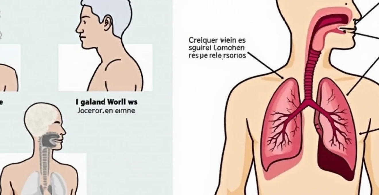
A persistent cough extending beyond two weeks often signals underlying respiratory conditions that require careful evaluation and targeted treatment. While acute coughs typically resolve within seven to ten days following viral upper respiratory infections, prolonged coughing episodes frequently indicate more complex pathophysiological processes affecting the respiratory tract. Medical professionals classify persistent coughs lasting fourteen days or longer as subacute conditions, distinguishing them from acute episodes and chronic manifestations that extend beyond eight weeks. Understanding the clinical significance of two-week cough duration becomes crucial for both healthcare providers and patients seeking appropriate therapeutic interventions. The diagnostic complexity surrounding persistent cough presentations necessitates comprehensive assessment protocols that consider multiple anatomical systems and potential underlying aetiologies affecting respiratory function.
Clinical definition and diagnostic criteria for persistent cough beyond fourteen days
Medical terminology defines subacute cough as symptoms persisting between three and eight weeks, with the two-week threshold representing a critical diagnostic milestone for healthcare practitioners. The International Classification of Diseases (ICD-11) categorises persistent cough based on duration, with fourteen-day presentations falling within the transitional period between acute and subacute classifications. Clinical guidelines established by respiratory medicine societies emphasise the importance of systematic evaluation when cough symptoms extend beyond the typical seven to ten-day resolution timeline expected following viral respiratory infections.
Diagnostic criteria for persistent cough assessment encompass multiple clinical parameters including symptom character, associated manifestations, patient demographics, and environmental exposure history. Healthcare providers utilise structured assessment protocols incorporating cough frequency measurements, sputum production analysis, and accompanying respiratory symptoms to establish preliminary diagnostic frameworks. The temporal relationship between initial symptom onset and current presentation provides valuable insights into potential underlying pathological processes affecting respiratory tract function.
Comprehensive diagnostic evaluation requires consideration of patient-specific factors including age, smoking history, occupational exposures, and pre-existing medical conditions that may influence cough persistence. Laboratory investigations, radiological imaging, and specialised pulmonary function testing contribute to differential diagnosis formulation when clinical presentations suggest underlying respiratory pathology. Modern diagnostic approaches emphasise evidence-based assessment protocols that optimise resource utilisation while ensuring thorough evaluation of persistent cough manifestations.
Upper respiratory tract infections: Post-Viral syndrome and rhinosinusitis complications
Post-infectious cough following rhinovirus and influenza A infections
Post-viral cough syndrome represents one of the most common aetiologies underlying persistent respiratory symptoms extending beyond the typical acute infection resolution period. Rhinovirus infections, accounting for approximately 30-50% of common cold episodes, frequently result in prolonged cough manifestations due to sustained inflammatory responses affecting respiratory epithelium. The pathophysiological mechanism involves viral-induced damage to ciliary function and increased mucus production, creating conditions conducive to persistent cough reflex activation. Inflammatory mediator release following initial viral infection continues affecting respiratory tract sensitivity for weeks beyond viral clearance, explaining symptom persistence despite resolution of systemic illness manifestations.
Influenza A virus infections demonstrate particular propensity for generating prolonged respiratory symptoms, with cough persistence rates approaching 25-40% of affected individuals experiencing symptoms beyond fourteen days. The neuraminidase activity characteristic of influenza viruses contributes to extensive respiratory epithelial damage, requiring substantial regeneration periods for complete functional recovery. Clinical presentations typically involve dry, non-productive cough patterns that may evolve into productive manifestations as epithelial repair processes progress and normal mucociliary function gradually restores.
Chronic rhinosinusitis with nasal polyps and cough reflex hypersensitivity
Chronic rhinosinusitis complicated by nasal polyposis frequently manifests as persistent cough due to continuous postnasal drainage stimulating sensitive laryngeal and tracheal receptors. The inflammatory cascade involving eosinophilic infiltration and cytokine-mediated responses creates sustained mucus production that triggers repetitive cough episodes. Upper airway cough syndrome , previously termed postnasal drip syndrome, encompasses these mechanistic pathways linking sino-nasal pathology with persistent lower respiratory tract symptoms.
Cough reflex hypersensitivity develops secondary to chronic inflammation affecting vagal nerve pathways responsible for respiratory protection mechanisms. This neuroplasticity phenomenon results in enhanced sensitivity to normally benign stimuli, perpetuating cough responses beyond the initial inflammatory trigger. Treatment approaches targeting both sino-nasal inflammation and cough receptor sensitivity demonstrate superior efficacy compared to single-modality therapeutic interventions.
Bacterial superinfection: streptococcus pneumoniae and haemophilus influenzae
Secondary bacterial infections complicating initial viral respiratory episodes frequently contribute to persistent cough manifestations extending beyond the expected recovery timeframe. Streptococcus pneumoniae represents the most common bacterial pathogen causing post-viral pneumonia, with clinical presentations ranging from subtle respiratory symptoms to frank pneumonic consolidation. The diagnostic challenge involves distinguishing bacterial superinfection from prolonged viral syndrome, requiring careful assessment of symptom progression patterns and inflammatory marker evaluation.
Haemophilus influenzae colonisation often occurs following viral-induced respiratory epithelial damage, creating opportunities for bacterial adherence and subsequent infection development. This opportunistic pathogen particularly affects individuals with underlying chronic respiratory conditions or compromised immune function. Clinical deterioration following initial improvement suggests bacterial superinfection requiring targeted antimicrobial therapy based on local resistance patterns and patient-specific risk factors.
Mycoplasma pneumoniae atypical pneumonia presentation
Mycoplasma pneumoniae infections frequently present with insidious onset patterns and prolonged symptom courses that extend well beyond typical bacterial pneumonia timelines. This atypical pathogen demonstrates particular affinity for respiratory epithelium, causing sustained inflammatory responses that manifest as persistent, non-productive cough lasting several weeks. The lack of cell wall structures in mycoplasma organisms necessitates specific antimicrobial selections, with macrolide antibiotics representing first-line therapeutic options for confirmed infections.
Diagnostic confirmation of mycoplasma pneumoniae infection relies on serological testing or polymerase chain reaction techniques, as traditional bacterial culture methods prove ineffective for this fastidious organism. Clinical presentations often include gradual symptom onset, constitutional symptoms, and radiological findings disproportionate to physical examination findings. The persistent cough associated with mycoplasma infections may continue for 4-6 weeks despite appropriate antimicrobial therapy, reflecting the organism’s impact on respiratory epithelial function and repair processes.
Lower respiratory pathology: bronchial and pulmonary manifestations
Acute bronchitis progression to chronic obstructive patterns
Acute bronchitis episodes occasionally progress toward chronic obstructive patterns, particularly in individuals with pre-existing respiratory vulnerabilities or significant smoking histories. The inflammatory cascade affecting bronchial epithelium may persist beyond typical acute episode resolution, creating sustained airway hyperresponsiveness and mucus hypersecretion. This progression involves complex interactions between inflammatory mediators, epithelial repair mechanisms, and environmental exposure factors that influence recovery trajectories. Bronchial wall remodelling processes initiated during acute inflammatory phases may continue for weeks, explaining persistent cough manifestations and associated respiratory symptoms.
Clinical assessment of potential progression requires careful monitoring of symptom patterns, spirometry measurements, and response to bronchodilator therapy. Patients demonstrating persistent airflow limitation or continued inflammatory markers beyond expected recovery timelines warrant comprehensive evaluation for underlying chronic obstructive pulmonary disease development. Early identification and intervention may prevent progression to more severe obstructive patterns requiring long-term therapeutic management strategies.
Community-acquired pneumonia with delayed resolution
Community-acquired pneumonia presentations occasionally demonstrate delayed resolution patterns, with persistent cough representing a common residual symptom extending beyond radiological clearance. The pathophysiological basis involves sustained inflammatory responses affecting alveolar and bronchial structures, requiring extended periods for complete functional recovery. Pneumonic consolidation resolution typically precedes symptom resolution by several weeks, creating diagnostic challenges when evaluating persistent respiratory complaints following pneumonia treatment.
Risk factors for delayed pneumonia resolution include advanced age, comorbid conditions, initial severity of presentation, and specific pathogen characteristics. Streptococcus pneumoniae and Legionella pneumophila demonstrate particular associations with prolonged recovery periods and persistent cough manifestations. Treatment completion does not guarantee immediate symptom resolution, with cough persistence lasting 2-6 weeks representing normal recovery patterns in many cases.
Bronchiectasis exacerbation and pseudomonas aeruginosa colonisation
Bronchiectasis exacerbations frequently present with increased cough production, purulent sputum characteristics, and systemic inflammatory responses that may persist for extended periods despite appropriate antimicrobial therapy. The structural airway damage characteristic of bronchiectasis creates persistent reservoirs for bacterial colonisation, particularly with Pseudomonas aeruginosa organisms demonstrating high-level antibiotic resistance. These chronic infections contribute to ongoing inflammatory responses that manifest as persistent productive cough with characteristic purulent sputum production.
Pseudomonas colonisation represents a significant complication in bronchiectasis management, requiring specialised antimicrobial approaches including inhaled antibiotic formulations and extended treatment durations. The biofilm formation capacity of these organisms creates additional therapeutic challenges, as standard antibiotic concentrations may prove insufficient for eradication. Airway clearance techniques combined with targeted antimicrobial therapy represent essential components of comprehensive bronchiectasis exacerbation management protocols.
Interstitial lung disease: idiopathic pulmonary fibrosis early indicators
Early-stage idiopathic pulmonary fibrosis frequently presents with insidious dry cough development that may initially be attributed to more common respiratory conditions. The pathogenesis involves progressive fibroblast proliferation and collagen deposition affecting alveolar architecture, creating mechanical stimuli that trigger persistent cough reflexes. This progressive fibrotic process typically demonstrates gradual symptom onset over months to years, though acute exacerbations may precipitate more rapid symptom development requiring urgent evaluation and intervention.
Diagnostic challenges arise due to non-specific early presentations that overlap with more common respiratory conditions, potentially delaying appropriate specialist referral and treatment initiation. High-resolution computed tomography represents the gold standard imaging modality for detecting early fibrotic changes, while pulmonary function testing reveals characteristic restrictive patterns with reduced diffusion capacity. Early recognition becomes crucial for initiating antifibrotic therapies that may slow disease progression and preserve respiratory function.
Asthma variants and allergic respiratory syndromes
Cough-variant asthma and methacholine challenge testing
Cough-variant asthma represents a distinct phenotype characterised by chronic cough as the primary or sole manifestation, without typical wheezing or dyspnoea symptoms associated with classic asthma presentations. This condition affects approximately 10-15% of individuals with asthma, creating diagnostic challenges due to the absence of characteristic bronchospastic symptoms. The underlying pathophysiology involves airway hyperresponsiveness affecting cough receptors without significant bronchial smooth muscle contraction, explaining the isolated cough manifestations. Inflammatory mediator release in response to typical asthma triggers creates sustained cough receptor sensitisation that persists between exposure episodes.
Methacholine challenge testing provides valuable diagnostic information for confirming airway hyperresponsiveness in patients with isolated chronic cough. This provocation test measures bronchial responsiveness to increasing concentrations of methacholine, with positive results supporting asthma diagnosis even in the absence of spontaneous bronchospasm. The test protocol requires careful monitoring and immediate bronchodilator availability due to potential for inducing significant airway constriction in susceptible individuals.
Occupational asthma from isocyanate and flour dust exposure
Occupational asthma development following workplace exposure to isocyanates or flour dust frequently manifests with persistent cough symptoms that may continue for weeks or months after initial sensitisation episodes. Isocyanate exposure, common in automotive, construction, and manufacturing industries, triggers IgE-mediated inflammatory responses that create sustained airway hyperresponsiveness. The temporal relationship between workplace exposure and symptom development provides crucial diagnostic information, with symptoms typically improving during weekends or vacation periods in early disease stages.
Flour dust sensitisation affects approximately 5-10% of bakery workers and food processing employees, creating occupational asthma patterns characterised by persistent cough, particularly during work shifts. The allergenic proteins present in wheat, rye, and other grain flours trigger respiratory sensitisation that may progress from mild symptoms to severe asthmatic responses without appropriate exposure control measures. Diagnostic confirmation requires specific IgE testing and potentially controlled exposure challenges conducted in specialised occupational health facilities.
Allergic bronchopulmonary aspergillosis in atopic patients
Allergic bronchopulmonary aspergillosis (ABPA) represents a complex hypersensitivity reaction to Aspergillus fumigatus occurring predominantly in individuals with underlying asthma or cystic fibrosis. The condition manifests with persistent productive cough, often containing brownish sputum plugs, along with recurrent pneumonic infiltrates and elevated specific IgE levels. The pathophysiology involves both immediate hypersensitivity reactions and delayed inflammatory responses that create sustained airway inflammation and mucus impaction.
Diagnostic criteria for ABPA include clinical symptoms, radiological findings, immunological parameters, and mycological evidence of Aspergillus sensitisation. Central bronchiectasis development represents a characteristic feature distinguishing ABPA from other allergic respiratory conditions, requiring high-resolution computed tomography for accurate assessment. Treatment protocols typically involve corticosteroid therapy for inflammatory control and antifungal medications for Aspergillus suppression, though complete eradication remains challenging due to the widespread environmental presence of these organisms.
Eosinophilic bronchitis without airway hyperresponsiveness
Eosinophilic bronchitis presents as chronic cough without associated airway hyperresponsiveness or variable airflow obstruction characteristic of asthma. This condition involves eosinophilic inflammatory infiltration of bronchial mucosa and submucosa, creating cough receptor stimulation without significant bronchial smooth muscle involvement. The diagnostic distinction from cough-variant asthma requires comprehensive evaluation including methacholine challenge testing to confirm absence of airway hyperresponsiveness alongside eosinophilic inflammation documentation through induced sputum analysis.
Treatment approaches for eosinophilic bronchitis focus primarily on anti-inflammatory therapy using inhaled corticosteroids, which demonstrate excellent efficacy for symptom control in most patients. The condition typically responds rapidly to appropriate anti-inflammatory treatment, with cough resolution occurring within 2-4 weeks of corticosteroid initiation. Long-term management may require sustained low-dose inhaled corticosteroid therapy to prevent symptom recurrence, though some patients achieve sustained remission following initial treatment courses.
Gastroesophageal and Cardiac-Related cough mechanisms
Gastroesophageal reflux disease (GERD) represents a frequently overlooked cause of persistent cough, affecting approximately 25-40% of individuals presenting with chronic respiratory symptoms. The pathophysiological mechanism involves acid reflux-induced stimulation of vagal afferent fibres in the distal oesophagus, creating reflex cough responses without direct gastric acid contact with respiratory structures. This neurally-mediated pathway explains why traditional antacid therapy may prove insufficient for cough control, requiring comprehensive acid suppression strategies targeting both volume and acidity of refluxed material. Microaspiration of gastric contents may also contribute to direct respiratory tract irritation, particularly during supine positioning when gravitational factors favour retrograde flow.
The diagnostic challenge surrounding GERD-related cough involves the frequent absence of classic heartburn symptoms, with up to 75% of patients demonstrating isolated respiratory manifestations. Ambulatory pH monitoring provides objective evidence of acid reflux episodes, though normal results do not exclude GERD as a contributing factor due to non-acid reflux contributions to symptom generation. The temporal relationship between meals, positioning changes, and cough episodes offers valuable diagnostic clues supporting reflux-related aetiology.
Cardiac-related cough mechanisms involve multiple pathophysiological pathways including pulmonary venous congestion, increased left atrial pressure, and activation of pulmonary stretch receptors. Heart failure patients frequently develop persistent dry cough due to interstitial pulmonary oedema and increased lung stiffness affecting mechanoreceptor function. The cough typically worsens in supine positions and may be accompanied by orthopnoea, paroxysmal nocturnal dyspnoea, and exercise intolerance. Angiotensin-converting enzyme (ACE) inhibitor
-related cough represents another significant consideration, affecting approximately 10-15% of patients prescribed these medications for cardiovascular protection. The mechanism involves bradykinin accumulation due to reduced enzymatic degradation, leading to increased cough receptor sensitivity and dry cough development that may persist for weeks after medication initiation.
Malignancy screening: lung cancer and metastatic disease indicators
Persistent cough extending beyond two weeks warrants careful consideration of underlying malignancy, particularly in high-risk populations including current or former smokers, individuals with occupational carcinogen exposures, and patients over 50 years of age with new-onset respiratory symptoms. Primary lung cancer represents the most concerning malignant cause of chronic cough, with adenocarcinoma and squamous cell carcinoma demonstrating particular associations with persistent respiratory symptoms. The pathophysiological mechanism involves tumour-related airway obstruction, inflammatory mediator release, and direct irritation of bronchial structures by malignant tissue growth. Early detection through appropriate screening protocols significantly impacts treatment outcomes and survival rates for patients with lung malignancies.
Clinical presentation patterns may include subtle changes in cough character, hemoptysis development, unintentional weight loss, and progressive dyspnoea that distinguishes malignant processes from benign inflammatory conditions. The diagnostic approach requires systematic evaluation incorporating patient risk stratification, comprehensive imaging studies, and potentially invasive procedures for tissue confirmation when clinical suspicion remains elevated. Low-dose computed tomography screening demonstrates proven efficacy for detecting early-stage lung cancers in high-risk populations, though false-positive rates necessitate careful patient selection and multidisciplinary management approaches.
Metastatic disease to pulmonary structures may present with persistent cough symptoms, particularly when primary malignancies originate from breast, colorectal, renal, or melanoma sources. The pattern of pulmonary metastases influences symptom development, with multiple small nodules typically causing less dramatic presentations compared to larger masses or lymphangitic spread patterns. Tumour emboli within pulmonary vasculature create additional pathophysiological mechanisms contributing to cough development through vascular occlusion and inflammatory responses affecting surrounding lung tissue.
Haematological malignancies including lymphomas and leukaemias occasionally present with persistent respiratory symptoms due to mediastinal lymphadenopathy, pulmonary infiltration, or opportunistic infections secondary to immunosuppression. The diagnostic challenge involves distinguishing primary haematological involvement from secondary infectious complications that frequently complicate these conditions. Comprehensive evaluation requires integration of imaging findings, laboratory parameters including complete blood counts and lactate dehydrogenase levels, and potentially bone marrow assessment when systemic involvement is suspected.
The temporal relationship between symptom onset and progression provides valuable diagnostic information, with malignant processes typically demonstrating gradual symptom escalation over weeks to months rather than the fluctuating patterns characteristic of inflammatory conditions. Constitutional symptoms including unexplained fever, night sweats, and progressive fatigue accompanying persistent cough warrant expedited evaluation for potential malignant aetiologies. Red flag symptoms requiring immediate specialist referral include hemoptysis, significant weight loss exceeding 5% of body weight, persistent hoarseness suggesting recurrent laryngeal nerve involvement, and superior vena cava syndrome manifestations indicating mediastinal mass effects.
Diagnostic imaging protocols for malignancy screening typically commence with chest radiography, though normal results do not exclude significant pathology given the limited sensitivity for detecting early-stage malignancies or small pulmonary nodules. High-resolution computed tomography provides superior sensitivity for detecting pulmonary masses, lymphadenopathy, and pleural abnormalities that may contribute to persistent cough symptoms. Additional imaging modalities including positron emission tomography may be indicated when initial studies suggest malignant processes requiring staging evaluation and treatment planning.
The integration of clinical assessment, imaging findings, and tissue sampling when appropriate enables accurate diagnosis and staging of malignant conditions presenting with persistent cough. Bronchoscopy with bronchoalveolar lavage and transbronchial biopsy provides direct visualisation of endobronchial abnormalities while enabling tissue acquisition for histological confirmation. Advanced bronchoscopic techniques including endobronchial ultrasound and electromagnetic navigation enhance diagnostic yield for peripheral pulmonary lesions and mediastinal lymph node sampling.
Treatment considerations for malignancy-related cough involve addressing both the underlying neoplastic process and symptomatic management of respiratory complaints. Antitussive therapies may provide temporary symptom relief while definitive oncological treatments including surgery, chemotherapy, and radiation therapy target the primary pathological process. Palliative care integration becomes essential for patients with advanced malignancies, focusing on symptom control and quality of life optimisation through comprehensive supportive care approaches including cough suppression, bronchodilator therapy, and psychosocial support services.
The prognosis for malignancy-related persistent cough varies significantly based on tumour type, staging, patient performance status, and response to treatment interventions. Early-stage lung cancers detected through systematic evaluation of persistent cough symptoms demonstrate significantly improved survival outcomes compared to advanced presentations, emphasising the importance of prompt evaluation and appropriate specialist referral when clinical presentations suggest potential malignant aetiologies. Long-term survivorship considerations include ongoing surveillance for disease recurrence and management of treatment-related complications that may affect respiratory function and cough symptom development.