Numbness in the ring and little fingers represents one of the most diagnostically challenging presentations in peripheral nerve disorders. Unlike the more commonly recognised carpal tunnel syndrome affecting the thumb and first three fingers, numbness affecting the fourth and fifth digits often signals involvement of the ulnar nerve pathway or cervical nerve root compression. This specific pattern of sensory loss can significantly impact daily activities, from typing and gripping objects to performing fine motor tasks that require precise finger coordination.
The anatomical complexity of nerve pathways serving the last two fingers creates multiple potential sites of compression or injury. Understanding these pathways becomes crucial for healthcare professionals and patients alike, as early recognition and appropriate intervention can prevent permanent nerve damage and restore normal function. The differential diagnosis between various causes requires systematic evaluation of symptoms, anatomical knowledge, and appropriate diagnostic testing.
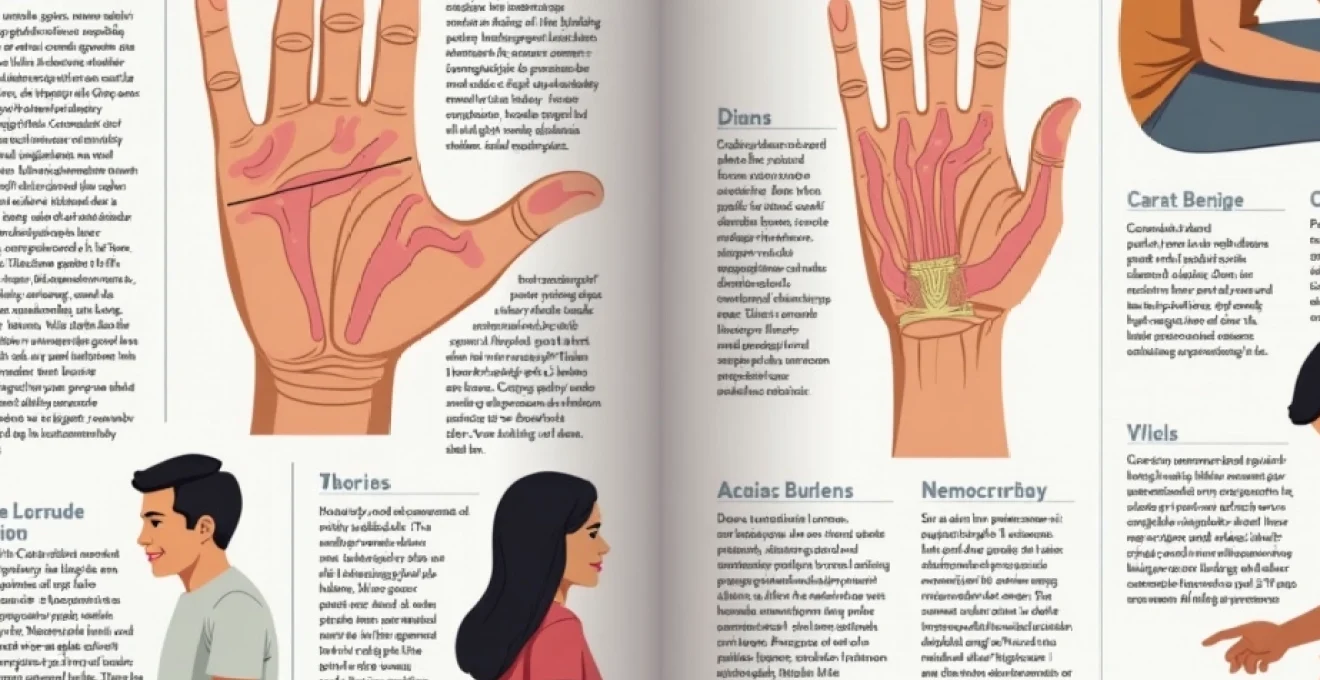
Ulnar nerve entrapment: primary cause of ring and little finger numbness
The ulnar nerve serves as the primary neural supply for sensation in the ring and little fingers, making its entrapment the most frequent cause of numbness in these digits. This nerve originates from the C8 and T1 nerve roots in the neck, travels through the brachial plexus, and descends along the medial aspect of the arm before passing through specific anatomical tunnels where compression commonly occurs. Ulnar nerve entrapment accounts for approximately 25% of all peripheral nerve compression syndromes, second only to carpal tunnel syndrome in frequency.
The clinical presentation of ulnar nerve entrapment typically follows a predictable pattern, beginning with intermittent numbness and tingling in the ring and little fingers. Patients often describe the sensation as “pins and needles” or a “dead feeling” that initially occurs during specific activities or positions. As the condition progresses, the numbness becomes more persistent and may be accompanied by weakness in grip strength, particularly affecting the ability to pinch objects between the thumb and index finger.
Cubital tunnel syndrome pathophysiology and compression mechanisms
Cubital tunnel syndrome represents the most common site of ulnar nerve entrapment, occurring at the elbow where the nerve passes through a narrow fibro-osseous tunnel behind the medial epicondyle. The cubital tunnel is formed by the medial epicondyle, olecranon process, and the arcuate ligament, creating a confined space that becomes even more restricted during elbow flexion. Anatomical studies have demonstrated that the tunnel volume decreases by up to 55% when the elbow is flexed beyond 90 degrees, explaining why symptoms often worsen during sleep or prolonged elbow flexion.
The pathophysiology involves mechanical compression leading to nerve ischaemia and subsequent demyelination. Prolonged compression results in intraneural oedema, which further increases pressure within the tunnel, creating a cycle of progressive nerve dysfunction. Occupational factors such as prolonged computer use, repetitive elbow flexion, or direct pressure on the elbow can precipitate or exacerbate the condition.
Guyon’s canal syndrome: ulnar nerve compression at the wrist
Guyon’s canal syndrome occurs when the ulnar nerve becomes compressed as it passes through the ulnar tunnel at the wrist. This tunnel, bounded by the hook of the hamate bone and the pisiform bone, represents the second most common site of ulnar nerve entrapment. The syndrome presents unique challenges in diagnosis as the sensory and motor symptoms can vary depending on the exact location of compression within the canal.
The canal can be divided into three zones, each associated with different symptom patterns. Zone 1 compression affects both motor and sensory branches, causing numbness in the ring and little fingers along with weakness in intrinsic hand muscles. Zone 2 compression primarily affects the deep motor branch, resulting in weakness without significant sensory loss. Zone 3 compression involves the superficial sensory branch, causing numbness without motor involvement.
Anatomical variations in ulnar nerve distribution patterns
Individual variations in ulnar nerve anatomy significantly influence the presentation of numbness patterns in the last two fingers. The classical description suggests complete sensory supply to the little finger and the ulnar half of the ring finger, but anatomical studies reveal considerable variation in these patterns. Approximately 15-20% of individuals demonstrate variant innervation patterns where the median nerve contributes to ring finger sensation, potentially leading to atypical symptom presentations.
These variations become clinically relevant when evaluating patients with finger numbness, as they can lead to diagnostic confusion and inappropriate treatment decisions. Understanding these patterns helps explain why some patients with confirmed ulnar nerve entrapment may not present with the classical numbness distribution, and why comprehensive nerve testing becomes essential for accurate diagnosis.
Differential diagnosis between C8 radiculopathy and ulnar neuropathy
Distinguishing between C8 radiculopathy and ulnar neuropathy presents a common diagnostic challenge, as both conditions can cause numbness in the ring and little fingers. The key differentiating factors lie in the distribution of symptoms and associated findings. C8 radiculopathy typically affects the entire C8 dermatome, extending beyond the ulnar nerve distribution to include the medial forearm and potentially the medial aspect of the middle finger.
Clinical examination reveals additional distinguishing features. Ulnar neuropathy primarily affects intrinsic hand muscles innervated by the ulnar nerve, while C8 radiculopathy may involve muscles innervated by multiple nerves arising from the C8 root, including some median nerve-innervated muscles. Provocative testing such as Tinel’s sign at the elbow or wrist suggests peripheral nerve entrapment, while cervical spine manoeuvres that reproduce symptoms point toward radiculopathy.
Cervical radiculopathy: C8 nerve root involvement and digital sensory loss
Cervical radiculopathy affecting the C8 nerve root represents a significant cause of numbness in the last two fingers, often mimicking peripheral ulnar nerve entrapment. The C8 nerve root contributes to both the median and ulnar nerves through the brachial plexus, making its compression a potential source of complex symptom patterns affecting multiple finger distributions. Epidemiological data suggests that C8 radiculopathy accounts for approximately 10-15% of all cervical radiculopathies, though it remains underdiagnosed due to its similarity to more common peripheral nerve conditions.
The pathophysiology involves compression or irritation of the C8 nerve root as it exits the spinal canal, typically between the C7 and T1 vertebrae. This compression can result from disc herniation, osteophyte formation, foraminal stenosis, or inflammatory conditions affecting the nerve root. The resulting symptoms extend beyond simple numbness to include pain, weakness, and reflex changes that follow the C8 dermatome and myotome distribution.
C8 dermatome distribution and clinical presentation patterns
The C8 dermatome encompasses the ring and little fingers, the ulnar aspect of the hand, and extends proximally along the medial forearm to the medial elbow region. This distribution explains why patients with C8 radiculopathy often experience numbness that extends well beyond the fingers to include forearm symptoms. The sensory loss typically follows a distinct pattern that can be mapped during clinical examination, helping to differentiate it from peripheral nerve entrapment syndromes.
Clinical presentation varies depending on the severity and chronicity of nerve root compression. Acute presentations may involve sharp, shooting pain that radiates from the neck down the medial arm to the fingers, often accompanied by numbness and tingling. Chronic presentations tend to manifest as persistent numbness with associated weakness in C8-innervated muscles, including some intrinsic hand muscles and wrist flexors.
Cervical disc herniation impact on lower trunk brachial plexus
Disc herniation at the C7-T1 level can significantly impact the lower trunk of the brachial plexus, affecting both C8 and T1 nerve roots. This anatomical relationship means that disc pathology at this level can produce complex symptom patterns affecting multiple nerve distributions simultaneously. The herniated disc material can compress nerve roots directly or create inflammatory responses that affect neural function over extended periods.
The mechanical compression varies with neck position, explaining why patients often report symptom fluctuation based on head and neck positioning. Extension and ipsilateral rotation typically worsen symptoms by further narrowing the already compromised neural foramen, while neck flexion and contralateral rotation may provide temporary relief by opening the foramen and reducing compression.
Spurling’s test and provocative manoeuvres for C8 radiculopathy
Spurling’s test serves as a valuable clinical tool for identifying C8 radiculopathy, involving passive extension and ipsilateral rotation of the cervical spine while applying axial compression. A positive test reproduces the patient’s symptoms of numbness and pain radiating into the C8 distribution, suggesting nerve root compression. The test demonstrates high specificity for cervical radiculopathy, though sensitivity varies depending on the degree of nerve root compression.
Additional provocative manoeuvres include the cervical distraction test, where upward traction on the head and neck relieves symptoms by opening the neural foramina and reducing nerve root compression. The upper limb tension test specifically targeting the ulnar nerve can help differentiate between cervical and peripheral causes of symptoms by putting selective stress on different portions of the neural pathway.
MRI findings in C7-T1 intervertebral disc pathology
Magnetic resonance imaging provides crucial information about C7-T1 disc pathology and its impact on neural structures. High-resolution MRI can demonstrate disc herniation, foraminal stenosis, and nerve root compression with remarkable detail. T2-weighted images effectively show disc degeneration and herniation, while post-contrast studies can reveal inflammatory changes around compressed nerve roots.
The correlation between MRI findings and clinical symptoms requires careful interpretation, as asymptomatic disc abnormalities occur frequently in the general population. Clinically relevant findings include significant foraminal narrowing, nerve root displacement, and signal changes within the nerve root itself suggesting oedema or inflammation. The combination of appropriate clinical symptoms and corresponding MRI changes provides strong evidence for C8 radiculopathy as the cause of finger numbness.
Thoracic outlet syndrome: neurovascular bundle compression effects
Thoracic outlet syndrome represents a complex condition involving compression of the neurovascular bundle as it traverses the thoracic outlet, the space between the clavicle and first rib. While less common than other causes of finger numbness, thoracic outlet syndrome can produce distinctive patterns of sensory loss affecting the ring and little fingers, particularly when the lower trunk of the brachial plexus becomes compressed. The condition occurs in approximately 1 in 1000 individuals, with a higher prevalence in young, active adults and those performing overhead activities.
The thoracic outlet anatomy includes three potential sites of compression: the interscalene triangle, costoclavicular space, and subcoracoid space. Neurogenic thoracic outlet syndrome accounts for 95% of cases and typically involves compression of the lower trunk of the brachial plexus, which contains fibres from C8 and T1 nerve roots. This compression pattern explains why symptoms often mirror those seen in ulnar nerve distribution, creating diagnostic challenges.
Patients with thoracic outlet syndrome affecting the lower trunk typically experience numbness and tingling in the ring and little fingers that worsens with overhead arm positions or activities requiring prolonged arm elevation. The symptoms may be accompanied by aching pain in the shoulder and arm, and in some cases, patients report colour changes in the hand due to associated vascular compression. Provocative positioning such as abduction and external rotation of the arm often reproduces symptoms, distinguishing this condition from more distal nerve entrapments.
Diagnosis requires a combination of clinical history, physical examination findings, and exclusion of other conditions. The elevated arm stress test involves maintaining the arms in an abducted, externally rotated position for three minutes while opening and closing the hands. Reproduction of numbness and tingling, particularly in the ulnar distribution, suggests thoracic outlet syndrome. However, the diagnosis remains challenging due to the overlap of symptoms with other conditions and the lack of definitive diagnostic tests.
Diabetic peripheral neuropathy: metabolic impact on distal sensory fibres
Diabetic peripheral neuropathy affects approximately 50% of individuals with diabetes and represents one of the most common causes of distal sensory loss, including numbness in the ring and little fingers. The pathophysiology involves multiple metabolic pathways, including increased glucose flux through the polyol pathway, formation of advanced glycation end products, and oxidative stress leading to progressive nerve fibre damage. Distal sensorimotor polyneuropathy is the most common form, typically presenting with a stocking-and-glove distribution of sensory loss.
The progression of diabetic neuropathy follows a predictable pattern, beginning with the longest nerve fibres in the feet before advancing to affect the hands. However, individuals with poorly controlled diabetes or longstanding disease may develop hand symptoms relatively early in the disease course. The numbness typically affects all fingers symmetrically, though patients may notice symptoms in the ring and little fingers more prominently due to their increased sensitivity to positional changes.
Clinical presentation includes burning pain, tingling, and numbness that is often worse at night. The sensory loss typically begins distally and progresses proximally in a length-dependent fashion. Small fibre neuropathy may precede large fibre involvement, causing painful burning sensations before obvious numbness develops. The condition is often accompanied by autonomic neuropathy, leading to additional symptoms such as gastroparesis, cardiac arrhythmias, or orthostatic hypotension.
Diagnosis relies on clinical presentation, nerve conduction studies, and quantitative sensory testing. Haemoglobin A1c levels provide information about glucose control over the preceding 2-3 months, with levels above 7% associated with increased neuropathy risk. Early detection and aggressive glucose control can slow progression, though established nerve damage is typically irreversible. Treatment focuses on glucose optimisation, pain management, and prevention of complications such as foot ulcers.
Carpal tunnel syndrome: median nerve involvement in fourth and fifth digit symptoms
While carpal tunnel syndrome classically affects the thumb, index, and middle fingers through median nerve compression, anatomical variations and severe compression can extend symptoms to include the ring finger. The median nerve provides sensation to the radial half of the ring finger in most individuals, though significant anatomical variation exists in this distribution. Advanced carpal tunnel syndrome can produce more extensive sensory changes, occasionally affecting portions of the ring finger and creating diagnostic confusion with ulnar nerve pathology.
The pathophysiology involves compression of the median nerve as it passes through the carpal tunnel, a rigid anatomical space bounded by the carpal bones and transverse carpal ligament. Increased pressure within this tunnel, whether from inflammation, fluid retention, or structural abnormalities, leads to nerve ischaemia and progressive dysfunction. The condition affects approximately 3-6% of the adult population, with higher rates in middle-aged women and individuals performing repetitive hand activities.
Atypical presentations of carpal tunnel syndrome may include numbness extending beyond the typical median nerve distribution, particularly when severe compression leads to extensive nerve dysfunction. Some patients report symptoms in the little finger, though this more commonly results from concurrent ulnar nerve pathology rather than isolated median nerve compression. Double crush syndrome describes the coexistence of proximal and distal nerve compression, where cervical radiculopathy and carpal tunnel syndrome occur simultaneously.
Electrodiagnostic testing remains the gold standard for confirming carpal tunnel syndrome, demonstrating prolonged distal motor latency and reduced sensory conduction velocity across the carpal tunnel. Treatment options range from conservative management with splinting and activity modification to surgical carpal tunnel release for severe or refractory cases. The prognosis is generally excellent with appropriate treatment, though outcomes depend on the duration and severity of compression before intervention.
Clinical diagnostic protocols: electromyography and nerve conduction studies
Electromyography and nerve conduction studies provide objective assessment of peripheral nerve function and serve as the diagnostic gold standard for evaluating numbness in the ring and little fingers. These studies can localise the site of nerve pathology, determine the severity of nerve dysfunction, and differentiate between various causes of finger numbness. Nerve conduction velocity measurements assess the speed of electrical impulse transmission along nerve fibres, while EMG evaluates the electrical activity of muscles innervated by suspect nerves.
Ulnar nerve conduction studies typically
include motor nerve conduction across the elbow and wrist, sensory nerve conduction studies, and late response testing including F-waves and H-reflexes. Abnormal findings in ulnar motor conduction studies may demonstrate prolonged distal motor latency, reduced conduction velocity across compression sites, or decreased compound muscle action potential amplitude indicating axonal loss. Sensory studies often show reduced or absent sensory nerve action potentials in the ring and little finger distributions.
Needle electromyography complements nerve conduction studies by evaluating muscle activity in ulnar-innervated muscles. The examination typically includes first dorsal interosseous, abductor digiti minimi, and flexor carpi ulnaris muscles to localise the level of ulnar nerve pathology. Denervation changes such as fibrillation potentials and positive sharp waves indicate active nerve damage, while chronic changes show motor unit remodelling with increased amplitude and duration of motor unit potentials.
Median nerve studies become essential when evaluating ring finger numbness, as anatomical variations can result in median nerve contribution to ring finger sensation. The study protocol includes motor conduction to the thenar muscles, sensory conduction to digits 1-3, and comparison studies between median and ulnar nerves innervating the ring finger. Comparative testing helps identify the relative contribution of each nerve to ring finger symptoms and guides appropriate treatment decisions.
C8 nerve root evaluation requires specific attention to muscles innervated by the C8 segment but supplied by different peripheral nerves. EMG examination of cervical paraspinal muscles, along with testing of both median and ulnar-innervated muscles, can help differentiate radiculopathy from peripheral nerve entrapment. The presence of cervical paraspinal denervation strongly suggests nerve root pathology, while normal paraspinal muscles with peripheral nerve abnormalities point toward distal entrapment syndromes.
Interpretation of electrodiagnostic results requires correlation with clinical findings and understanding of normal anatomical variants. Mild abnormalities may represent subclinical nerve dysfunction, while severe changes indicate established nerve damage requiring urgent intervention. The timing of studies is crucial, as acute nerve injuries may not demonstrate electrodiagnostic abnormalities for 2-3 weeks following onset, necessitating repeat testing in cases of suspected acute nerve trauma or compression.